Arm

Last updated
Arm
Extended arm.jpg
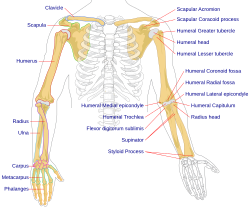
The left arm of a human male
Details
Artery Axillary artery
Vein Axillary vein
Nerve Brachial plexus
Identifiers
Latin brachium
Anatomical terminology

In human anatomy, the arm refers to the upper limb in common usage, although academically the term specifically means the upper arm [1] [2] between the glenohumeral joint (shoulder joint) and the elbow joint. The distal part of the upper limb between the elbow and the radiocarpal joint (wrist joint) is known as the forearm or "lower" arm, and the extremity beyond the wrist is the hand.

Contents

By anatomical definitions, the bones, ligaments and skeletal muscles of the shoulder girdle, as well as the axilla between them, is considered parts of the upper limb, and thus also components of the arm. The Latin term brachium, which serves as a root word for naming many anatomical structures, may refer to either the upper limb as a whole or to the upper arm on its own. [3] [4] [5]

Anatomy

Bones

Bones of the upper limbs, together with shoulder girdles, that compose the human arm. Human arm bones diagram.svg
Bones of the upper limbs, together with shoulder girdles, that compose the human arm.

The humerus is one of the three long bones of the arm. It joins with the scapula at the shoulder joint and with the other long bones of the arm, the ulna and radius at the elbow joint. [6] The elbow is a complex hinge joint between the end of the humerus and the ends of the radius and ulna. [7]

Muscles

The arm is divided by a fascial layer (known as lateral and medial intermuscular septa) separating the muscles into two osteofascial compartments: the anterior and the posterior compartments of the arm. The fascia merges with the periosteum (outer bone layer) of the humerus. [8]

The anterior compartment contains three muscles: biceps brachii, brachialis and coracobrachialis muscles. They are all innervated by the musculocutaneous nerve. The posterior compartment contains only the triceps brachii muscle, supplied by the radial nerve. [9] [10] [11]

Nerve supply

Cutaneous innervation of the right upper extremity. Gray812and814.PNG
Cutaneous innervation of the right upper extremity.

The musculocutaneous nerve, from C5, C6, C7, is the main supplier of muscles of the anterior compartment. It originates from the lateral cord of the brachial plexus of nerves. It pierces the coracobrachialis muscle and gives off branches to the muscle, as well as to brachialis and biceps brachii. It terminates as the anterior cutaneous nerve of the forearm.

The radial nerve, which is from the fifth cervical spinal nerve to the first thoracic spinal nerve, originates as the continuation of the posterior cord of the brachial plexus. This nerve enters the lower triangular space (an imaginary space bounded by, amongst others, the shaft of the humerus and the triceps brachii) of the arm and lies deep to the triceps brachii. Here it travels with the deep artery of the arm, which sits in the radial groove of the humerus. This fact is very important clinically as a fracture of the shaft of the bone here can cause lesions or even transections in the nerve.

Other nerves passing through give no supply to the arm. These include:

Blood supply

Main arteries of the arm. Gray1235.png
Main arteries of the arm.

The main artery in the arm is the brachial artery. This artery is a continuation of the axillary artery. The point at which the axillary becomes the brachial is distal to the lower border of teres major. The brachial artery gives off an unimportant branch, the deep artery of arm. This branching occurs just below the lower border of teres major.

The brachial artery continues to the cubital fossa in the anterior compartment of the arm. It travels in a plane between the biceps and triceps muscles, the same as the median nerve and basilic vein. It is accompanied by venae comitantes (accompanying veins). It gives branches to the muscles of the anterior compartment. The artery is in between the median nerve and the tendon of the biceps muscle in the cubital fossa. It then continues into the forearm.

The deep artery of the arm travels through the lower triangular space with the radial nerve. From here onwards it has an intimate relationship with the radial nerve. They are both found deep to the triceps muscle and are located on the spiral groove of the humerus. Therefore, fracture of the bone may not only lead to lesion of the radial nerve, but also haematoma of the internal structures of the arm. The artery then continues on to anastamose with the recurrent radial branch of the brachial artery, providing a diffuse blood supply for the elbow joint.

Veins

The veins of the arm carry blood from the extremities of the limb, as well as drain the arm itself. The two main veins are the basilic and the cephalic veins. There is a connecting vein between the two, the median cubital vein, which passes through the cubital fossa and is clinically important for venepuncture (withdrawing blood).

The basilic vein travels on the medial side of the arm and terminates at the level of the seventh rib.

The cephalic vein travels on the lateral side of the arm and terminates as the axillary vein. It passes through the deltopectoral triangle, a space between the deltoid and the pectoralis major muscles.

Society and culture

In Hindu, Buddhist and Egyptian iconography the symbol of the arm is used to illustrate the power of the sovereign. In Hindu tradition gods are depicted with several arms which carry specific symbols of their powers. It is believed that several arms depict omnipotence of gods. In popular culture Thakur did not have arms in the movie Sholay.

In West Africa, the Bambara use forearm to symbolize the spirit, which is a link between God and man.

Three North Koreans surrender to USS Manchester by raising their arms Three Korean Communists in a fishing boat are captured by the USS Manchester off the coast of Korea. - NARA - 520792.tif
Three North Koreans surrender to USS Manchester by raising their arms

Symbolic gestures of raising both hands signal surrender, appeals for mercy, and justice. [12]

The arm, as can be seen here, formed one component of hieroglyphs Abydos KL 04-04 n23.jpg
The arm, as can be seen here, formed one component of hieroglyphs

Clinical significance

The cubital fossa is clinically important for venepuncture and for blood pressure measurement. [13]

When the arm is fractured this may refer to a fracture of the humerus bone.

Veins on the arm may be taken when a coronary artery bypass graft is needed.

Other animals

In other animals, the term arm can also be used for homologous or analogous structures (such as one of the paired forelimbs of a four-legged animal or the arms of cephalopods, respectively). In anatomical usage, the term arm may [3] sometimes refer specifically to the segment between the shoulder and the elbow, [14] [15] while the segment between the elbow and wrist is the forearm. However, in common, literary, and historical usage, arm refers to the entire upper limb from shoulder to wrist. This article uses the former definition; see upper limb for the wider definition. [8]

In primates, the arm is adapted for precise positioning of the hand and thus assist in the hand's manipulative tasks. The ball and socket shoulder joint allows for movement of the arms in a wide circular plane, while the structure of the two forearm bones which can rotate around each other allows for additional range of motion at that level. [16]

Additional images

See also

Related Research Articles

<span class="mw-page-title-main">Humerus</span> Long bone of the upper arm

The humerus is a long bone in the arm that runs from the shoulder to the elbow. It connects the scapula and the two bones of the lower arm, the radius and ulna, and consists of three sections. The humeral upper extremity consists of a rounded head, a narrow neck, and two short processes. The body is cylindrical in its upper portion, and more prismatic below. The lower extremity consists of 2 epicondyles, 2 processes, and 3 fossae. As well as its true anatomical neck, the constriction below the greater and lesser tubercles of the humerus is referred to as its surgical neck due to its tendency to fracture, thus often becoming the focus of surgeons.

<span class="mw-page-title-main">Biceps</span> Muscle on the front of the upper arm

The biceps or biceps brachii is a large muscle that lies on the front of the upper arm between the shoulder and the elbow. Both heads of the muscle arise on the scapula and join to form a single muscle belly which is attached to the upper forearm. While the biceps crosses both the shoulder and elbow joints, its main function is at the elbow where it flexes the forearm and supinates the forearm. Both these movements are used when opening a bottle with a corkscrew: first biceps screws in the cork (supination), then it pulls the cork out (flexion).

<span class="mw-page-title-main">Brachioradialis</span> Muscle of the upper limb

The brachioradialis is a muscle of the forearm that flexes the forearm at the elbow. It is also capable of both pronation and supination, depending on the position of the forearm. It is attached to the distal styloid process of the radius by way of the brachioradialis tendon, and to the lateral supracondylar ridge of the humerus.

<span class="mw-page-title-main">Radial nerve</span> Nerve in the human body that supplies the posterior portion of the upper limb

The radial nerve is a nerve in the human body that supplies the posterior portion of the upper limb. It innervates the medial and lateral heads of the triceps brachii muscle of the arm, as well as all 12 muscles in the posterior osteofascial compartment of the forearm and the associated joints and overlying skin.

<span class="mw-page-title-main">Brachial artery</span> Large blood vessel

The brachial artery is the major blood vessel of the (upper) arm. It is the continuation of the axillary artery beyond the lower margin of teres major muscle. It continues down the ventral surface of the arm until it reaches the cubital fossa at the elbow. It then divides into the radial and ulnar arteries which run down the forearm. In some individuals, the bifurcation occurs much earlier and the ulnar and radial arteries extend through the upper arm. The pulse of the brachial artery is palpable on the anterior aspect of the elbow, medial to the tendon of the biceps, and, with the use of a stethoscope and sphygmomanometer, often used to measure the blood pressure.

<span class="mw-page-title-main">Median nerve</span> Nerve of the upper limb

The median nerve is a nerve in humans and other animals in the upper limb. It is one of the five main nerves originating from the brachial plexus.

<span class="mw-page-title-main">Brachialis muscle</span> Flexor muscle in the upper arm

The brachialis is a muscle in the upper arm that flexes the elbow. It lies beneath the biceps brachii, and makes up part of the floor of the region known as the cubital fossa. It originates from the anterior aspect of the distal humerus; it inserts onto the tuberosity of the ulna. It is innervated by the musculocutaneous nerve, and commonly also receives additional innervation from the radial nerve. The brachialis is the prime mover of elbow flexion generating about 50% more power than the biceps.

The forearm is the region of the upper limb between the elbow and the wrist. The term forearm is used in anatomy to distinguish it from the arm, a word which is used to describe the entire appendage of the upper limb, but which in anatomy, technically, means only the region of the upper arm, whereas the lower "arm" is called the forearm. It is homologous to the region of the leg that lies between the knee and the ankle joints, the crus.

<span class="mw-page-title-main">Ulnar nerve</span> Nerve which runs near the ulna bone

In human anatomy, the ulnar nerve is a nerve that runs near the ulna bone. The ulnar collateral ligament of elbow joint is in relation with the ulnar nerve. The nerve is the largest in the human body unprotected by muscle or bone, so injury is common. This nerve is directly connected to the little finger, and the adjacent half of the ring finger, innervating the palmar aspect of these fingers, including both front and back of the tips, perhaps as far back as the fingernail beds.

<span class="mw-page-title-main">Wrist drop</span> Medical condition

Wrist drop is a medical condition in which the wrist and the fingers cannot extend at the metacarpophalangeal joints. The wrist remains partially flexed due to an opposing action of flexor muscles of the forearm. As a result, the extensor muscles in the posterior compartment remain paralyzed.

<span class="mw-page-title-main">Upper limb</span> Consists of the arm, forearm, and hand

The upper limbs or upper extremities are the forelimbs of an upright-postured tetrapod vertebrate, extending from the scapulae and clavicles down to and including the digits, including all the musculatures and ligaments involved with the shoulder, elbow, wrist and knuckle joints. In humans, each upper limb is divided into the arm, forearm and hand, and is primarily used for climbing, lifting and manipulating objects.

<span class="mw-page-title-main">Musculocutaneous nerve</span> Nerve in the arm

The musculocutaneous nerve is a mixed branch of the lateral cord of the brachial plexus derived from cervical spinal nerves C5-C7. It arises opposite the lower border of the pectoralis major. It provides motor innervation to the muscles of the anterior compartment of the arm: the coracobrachialis, biceps brachii, and brachialis. It provides sensory innervation to the lateral forearm. It courses through the anterior part of the arm, terminating 2 cm above elbow; after passing the lateral edge of the tendon of biceps brachii it is becomes known as the lateral cutaneous nerve of the forearm.

<span class="mw-page-title-main">Triceps</span> Muscle on the back of the upper arm

The triceps, or triceps brachii, is a large muscle on the back of the upper limb of many vertebrates. It consists of 3 parts: the medial, lateral, and long head. It is the muscle principally responsible for extension of the elbow joint.

<span class="mw-page-title-main">Cubital fossa</span> The human elbow pit

The cubital fossa,chelidon, grivet or inside of elbow, is the area on the anterior side of the upper part between the arm and forearm of a human or other hormid animals. It lies anteriorly to the elbow when in standard anatomical position.

<span class="mw-page-title-main">Coracobrachialis muscle</span> Muscle of the upper arm

The coracobrachialis muscle is a muscle in the upper medial part of the arm. It is located within the anterior compartment of the arm. It originates from the coracoid process of the scapula; it inserts onto the middle of the medial aspect of the body of the humerus. It is innervated by the musculocutaneous nerve. It acts to adduct and flex the arm.

The pronator teres is a muscle that, along with the pronator quadratus, serves to pronate the forearm. It has two origins, at the medial humeral supracondylar ridge and the ulnar tuberosity, and inserts near the middle of the radius.

<span class="mw-page-title-main">Fascial compartments of arm</span> Anatomical compartments

The fascial compartments of arm refers to the specific anatomical term of the compartments within the upper segment of the upper limb of the body. The upper limb is divided into two segments, the arm and the forearm. Each of these segments is further divided into two compartments which are formed by deep fascia – tough connective tissue septa (walls). Each compartment encloses specific muscles and nerves.

<span class="mw-page-title-main">Axillary space</span>

The axillary spaces are anatomic spaces. through which axillary contents leave the axilla. They consist of the quadrangular space, triangular space, and triangular interval. It is bounded by teres major, teres minor, medial border of the humerus, and long head of triceps brachii.

<span class="mw-page-title-main">Outline of human anatomy</span> Overview of and topical guide to human anatomy

The following outline is provided as an overview of and topical guide to human anatomy:

<span class="mw-page-title-main">Elbow</span> Joint between the upper and lower parts of the arm

The elbow is the region between the upper arm and the forearm that surrounds the elbow joint. The elbow includes prominent landmarks such as the olecranon, the cubital fossa, and the lateral and the medial epicondyles of the humerus. The elbow joint is a hinge joint between the arm and the forearm; more specifically between the humerus in the upper arm and the radius and ulna in the forearm which allows the forearm and hand to be moved towards and away from the body. The term elbow is specifically used for humans and other primates, and in other vertebrates forelimb plus joint is used.

References

  1. Gray, Henry (1918). "The Humerus (Arm Bone)".
  2. Forro, Stephen D. (July 31, 2021). "Anatomy, Shoulder and Upper Limb, Arm Structure and Function". StatPearls. PMID   29939618.
  3. 1 2 "brachium – free dictionary" . Retrieved December 2, 2013.
  4. "Dictionary.com" . Retrieved December 2, 2013.
  5. Encyclopaedia britannica 2013.
  6. Sam Jacob (2007). Human Anatomy: A Clinically-Orientated Approach. Elsevier Health Sciences. p. 5. ISBN   978-0443103735 . Retrieved January 19, 2018.
  7. Doral, Mahmut Nedim; Karlsson, Jon, eds. (2015-03-04). Sports injuries : prevention, diagnosis, treatment and rehabilitation. Berlin. ISBN   9783642368011. OCLC   1111734654.{{cite book}}: CS1 maint: location missing publisher (link)
  8. 1 2 Drake, Richard L. (Richard Lee) (15 November 2015). Gray's anatomy for students. Vogl, Wayne; Mitchell, Adam W. M.; Gray, Henry (Third ed.). Philadelphia, PA. ISBN   9780702051319. OCLC   881508489.{{cite book}}: CS1 maint: location missing publisher (link)
  9. Moore, Keith L. (2013-02-13). Clinically oriented anatomy. Dalley, Arthur F., II,, Agur, A. M. R. (Seventh ed.). Philadelphia. ISBN   978-1451119459. OCLC   813301028.{{cite book}}: CS1 maint: location missing publisher (link)
  10. Standring, Susan, ed. (2016). Gray's anatomy : the anatomical basis of clinical practice (Forty-first ed.). [Philadelphia]. ISBN   9780702052309. OCLC   920806541.{{cite book}}: CS1 maint: location missing publisher (link)
  11. "Elbow and Forearm". Kenhub. Retrieved 2019-09-14.
  12. Tresidder, Jack (1997). The Hutchinson Dictionary of Symbols. London: Helicon. p. 16. ISBN   1-85986-059-1.
  13. "How To Draw Blood Like A Pro: Step-By-Step Guide". nurse.org. Retrieved 2019-09-14.
  14. "Arm". The Lecturio Medical Concept Library. 3 November 2020. Retrieved 2021-06-22.
  15. Arm at the U.S. National Library of Medicine Medical Subject Headings (MeSH)
  16. Preuschoft, Holger; Hohn, Bianca; Scherf, Heike; Schmidt, Manuela; Krause, Cornelia; Witzel, Ulrich (April 2010). "Functional Analysis of the Primate Shoulder". International Journal of Primatology. 31 (2): 301–320. doi:10.1007/s10764-010-9399-1. ISSN   0164-0291. PMC   2860095 . PMID   20495602.