Flexor pollicis brevis muscle | |
---|---|
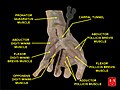
![]() Superficial muscles of the left hand, palmar view. | |
Details | |
Origin | Trapezium, flexor retinaculum |
Insertion | Thumb, proximal phalanx |
Artery | Superficial palmar arch |
Nerve | Recurrent branch of the median nerve, deep branch of ulnar nerve (medial head) |
Actions | Flexes the thumb at the first metacarpophalangeal joint |
Antagonist | Extensor pollicis longus and brevis |
Identifiers | |
Latin | musculus flexor pollicis brevis |
TA98 | A04.6.02.055 |
TA2 | 2522 |
FMA | 37378 |
Anatomical terms of muscle |
The flexor pollicis brevis is a muscle in the hand that flexes the thumb. It is one of three thenar muscles. [1] [2] It has both a superficial part and a deep part.
The muscle's superficial head arises from the distal edge of the flexor retinaculum and the tubercle of the trapezium, the most lateral bone in the distal row of carpal bones. [1] It passes along the radial side of the tendon of the flexor pollicis longus.
The deeper (and medial) head "varies in size and may be absent." [3] It arises from the trapezoid and capitate bones on the floor of the carpal tunnel, as well as the ligaments of the distal carpal row. [3]
Both heads become tendinous and insert together into the radial side of the base of the proximal phalanx of the thumb; [2] at the junction between the tendinous heads there is a sesamoid bone. [4]
The superficial head is usually innervated by the lateral terminal branch of the median nerve. [1] The deep part is often innervated by the deep branch of the ulnar nerve (C8, T1). [2] [3]
The flexor pollicis brevis receives its blood supply from the superficial palmar branches of radial artery. [5]
The flexor pollicis brevis flexes the thumb at the metacarpophalangeal joint, [1] as well as flexion and medial rotation of the 1st metacarpal bone at the carpometacarpal joint. [3]
Flexor pollicis brevis can, rarely, be completely absent at birth due to a congenital issue (as can the other muscles of the thenar eminence). [6]
The thumb is the first digit of the hand, next to the index finger. When a person is standing in the medical anatomical position, the thumb is the outermost digit. The Medical Latin English noun for thumb is pollex, and the corresponding adjective for thumb is pollical.
The radial nerve is a nerve in the human body that supplies the posterior portion of the upper limb. It innervates the medial and lateral heads of the triceps brachii muscle of the arm, as well as all 12 muscles in the posterior osteofascial compartment of the forearm and the associated joints and overlying skin.
The median nerve is a nerve in humans and other animals in the upper limb. It is one of the five main nerves originating from the brachial plexus.
The flexor digitorum profundus or flexor digitorum communis profundus is a muscle in the forearm of humans that flexes the fingers. It is considered an extrinsic hand muscle because it acts on the hand while its muscle belly is located in the forearm.
The thenar eminence is the mound formed at the base of the thumb on the palm of the hand by the intrinsic group of muscles of the thumb. The skin overlying this region is the area stimulated when trying to elicit a palmomental reflex. The word thenar comes from Ancient Greek θέναρ (thenar) 'palm of the hand'.
The ulnar nerve is a nerve that runs near the ulna, one of the two long bones in the forearm. The ulnar collateral ligament of elbow joint is in relation with the ulnar nerve. The nerve is the largest in the human body unprotected by muscle or bone, so injury is common. This nerve is directly connected to the little finger, and the adjacent half of the ring finger, innervating the palmar aspect of these fingers, including both front and back of the tips, perhaps as far back as the fingernail beds.
The trapezium bone is a carpal bone in the hand. It forms the radial border of the carpal tunnel.
The upper limbs or upper extremities are the forelimbs of an upright-postured tetrapod vertebrate, extending from the scapulae and clavicles down to and including the digits, including all the musculatures and ligaments involved with the shoulder, elbow, wrist and knuckle joints. In humans, each upper limb is divided into the shoulder, arm, elbow, forearm, wrist and hand, and is primarily used for climbing, lifting and manipulating objects. In anatomy, just as arm refers to the upper arm, leg refers to the lower leg.
The flexor pollicis longus is a muscle in the forearm and hand that flexes the thumb. It lies in the same plane as the flexor digitorum profundus. This muscle is unique to humans, being either rudimentary or absent in other primates. A meta-analysis indicated accessory flexor pollicis longus is present in around 48% of the population.
The palmaris longus is a muscle visible as a small tendon located between the flexor carpi radialis and the flexor carpi ulnaris, although it is not always present. Reviews report rates of absence in the general population ranging from 10–20%; however, the rate varies in different ethnic groups. Absence of the palmaris longus does not have an effect on grip strength. The lack of palmaris longus muscle does result in decreased pinch strength in fourth and fifth fingers. The absence of palmaris longus muscle is more prevalent in females than males.
In human anatomy, the adductor pollicis muscle is a muscle in the hand that functions to adduct the thumb. It has two heads: transverse and oblique.
In human anatomy, the palmar or volar interossei are four muscles, one on the thumb that is occasionally missing, and three small, unipennate, central muscles in the hand that lie between the metacarpal bones and are attached to the index, ring, and little fingers. They are smaller than the dorsal interossei of the hand.
The opponens pollicis is a small, triangular muscle in the hand, which functions to oppose the thumb. It is one of the three thenar muscles. It lies deep to the abductor pollicis brevis and lateral to the flexor pollicis brevis.
The palmar aponeurosis invests the muscles of the palm, and consists of central, lateral, and medial portions.
The deep palmar arch is an arterial network found in the palm. It is usually primarily formed from the terminal part of the radial artery. The ulnar artery also contributes through an anastomosis. This is in contrast to the superficial palmar arch, which is formed predominantly by the ulnar artery.
In the palm of the hand the median nerve is covered by the skin and the palmar aponeurosis, and rests on the tendons of the flexor muscles. Immediately after emerging from under the transverse carpal ligament the median nerve becomes enlarged and flattened and splits into a smaller, lateral, and a larger, medial portion.
The recurrent branch of the median nerve is the branch of the median nerve which supplies the thenar muscles. It is also occasionally referred to as the thenar branch of the median nerve, or the thenar muscular branch of the median nerve.
In the human body, the carpal tunnel or carpal canal is a flattened body cavity on the flexor (palmar/volar) side of the wrist, bounded by the carpal bones and flexor retinaculum. It forms the passageway that transmits the median nerve and the tendons of the extrinsic flexor muscles of the hand from the forearm to the hand. The median artery is an anatomical variant. When present it lies between the radial artery, and the ulnar artery and runs with the median nerve supplying the same structures innervated.
The muscles of the hand are the skeletal muscles responsible for the movement of the hand and fingers. The muscles of the hand can be subdivided into two groups: the extrinsic and intrinsic muscle groups. The extrinsic muscle groups are the long flexors and extensors. They are called extrinsic because the muscle belly is located on the forearm. The intrinsic group are the smaller muscles located within the hand itself. The muscles of the hand are innervated by the radial, median, and ulnar nerves from the brachial plexus.
The muscles of the thumb are nine skeletal muscles located in the hand and forearm. The muscles allow for flexion, extension, adduction, abduction and opposition of the thumb. The muscles acting on the thumb can be divided into two groups: The extrinsic hand muscles, with their muscle bellies located in the forearm, and the intrinsic hand muscles, with their muscles bellies located in the hand proper.
This article incorporates text in the public domain from page 461 of the 20th edition of Gray's Anatomy (1918)