Supraspinatus muscle

Last updated
Supraspinatus muscle
Supraspinatus muscle back4.png
Position of the supraspinatus muscle (red) seen from the back.
Supraspinatus.PNG
Posterior view of muscles connecting the upper extremity to the vertebral column. Supraspinatus muscle is labeled in red at right, while it is covered by other muscles at left.
Details
Origin Supraspinous fossa of scapula
Insertion Superior facet of greater tubercle of humerus
Artery Suprascapular artery
Nerve Suprascapular nerve
Actions Abduction of arm and stabilizes humerus
Identifiers
Latin musculus supraspinatus
TA98 A04.6.02.006
TA2 2457
FMA 9629
Anatomical terms of muscle

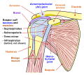
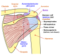
The supraspinatus (pl.: supraspinati) is a relatively small muscle of the upper back that runs from the supraspinous fossa superior portion of the scapula (shoulder blade) to the greater tubercle of the humerus. It is one of the four rotator cuff muscles and also abducts the arm at the shoulder. The spine of the scapula separates the supraspinatus muscle from the infraspinatus muscle, which originates below the spine.

Contents

Structure

Origin

The supraspinatus muscle arises from the medial two-thirds supraspinous fossa of the scapula. [1]

Insertion

The supraspinatus tendon inserts onto the superior facet of the greater tubercle of the humerus. [2]

Relations

The supraspinatus muscle tendon passes laterally beneath the cover of the acromion. [3] [4] The tendon blends with the shoulder joint capsule. [1]

Nerve supply

The supraspinatus muscle is innervated suprascapular nerve (C5-6) [1] of the upper trunk of the brachial plexus.

Function

The supraspinatus muscle performs abduction of the arm, and pulls the head of the humerus medially towards the glenoid cavity. [5] It independently prevents the head of the humerus from slipping inferiorly. [5] The supraspinatus works in cooperation with the deltoid muscle to perform abduction, including when the arm is in an adducted position. [5] Beyond 15 degrees, the deltoid muscle becomes increasingly more effective at abducting the arm and becomes the main propagator of this action. [6]

Clinical significance

The supraspinatus forms part of the rotator cuff and is one of its most frequently damaged components, whether from acute injury or gradual degeneration. [7] Bad posture and age are leading risk factors, with a high prevalence of unsymptomatic partial and full tears, as well as symptomatic syndromes with chronic pain. Connected pathologies include acromial impingement, frozen shoulder, and poor sleep, especially on the side. Both ultrasound and MRI are now effective methods of diagnosis.

Tear

Diagnosis

Antero-posterior projectional radiography of the shoulder may demonstrate a high-riding humeral head, with an acromiohumeral distance of less than 7 millimetres (0.28 in). [8]

Repair

One study has indicated that arthroscopic surgery for full-thickness supraspinatus tears is effective for improving shoulder functionality. [9]

A comparative effectiveness review of nonoperative and operative treatments for rotator cuff tears was performed at the University of Alberta Evidence-based Practice Center in 2010. The review identified one study which reported that, "Patients receiving early surgery had superior function compared with the delayed surgical group". The review noted that the level of significance of the study was not reported, and the review chose not to include it as one of their conclusions. Instead it concluded that "The paucity of evidence related to early versus delayed surgery is of particular concern, as patients and providers must decide whether to attempt initial nonoperative management or proceed immediately with surgical repair". In terms of operative techniques, differences in neither cuff integrity nor shoulder function were reported in studies comparing single-row versus double-row suture anchor fixation and mattress locking versus absorbable sutures. Postoperatively, a slight advantage was evident in patients who performed continuous passive motion alongside physical therapy, as opposed to those who solely performed physical therapy. There is insufficient evidence to adequately compare the effects of operative against nonoperative interventions. Complications were reported very seldom, or were not determined to be clinically significant. [10]

A 2016 study evaluating the effectiveness of arthroscopic treatment of rotator cuff calcification firmly supported surgical intervention. Calcification of the supraspinatus tendon is a major contributor to shoulder pain in the general population and is often worsened following a supraspinatus tear. The results of the study included the return to sports and original functionality of 95.8% of the patients after a mean of 5.3 post-operative months. A significant decrease in pain was observed over time following removal of the calcification. The study showed the overall effectiveness of arthroscopic procedures on shoulder repair, and the lack of risk experienced. [11] Before surgery, supraspinatus tendonitis should be ruled out as the cause of pain.

Paralysis

The suprascapular nerve which innervates the supraspinatus can be damaged along its course in fractures of the overlying clavicle, which can reduce the person's ability to initiate the abduction.[ citation needed ]

Related Research Articles

<span class="mw-page-title-main">Scapula</span> Bone that connects the humerus (upper arm bone) with the clavicle (collar bone)

The scapula, also known as the shoulder blade, is the bone that connects the humerus with the clavicle. Like their connected bones, the scapulae are paired, with each scapula on either side of the body being roughly a mirror image of the other. The name derives from the Classical Latin word for trowel or small shovel, which it was thought to resemble.

<span class="mw-page-title-main">Humerus</span> Long bone of the upper arm

The humerus is a long bone in the arm that runs from the shoulder to the elbow. It connects the scapula and the two bones of the lower arm, the radius and ulna, and consists of three sections. The humeral upper extremity consists of a rounded head, a narrow neck, and two short processes. The body is cylindrical in its upper portion, and more prismatic below. The lower extremity consists of 2 epicondyles, 2 processes, and 3 fossae. As well as its true anatomical neck, the constriction below the greater and lesser tubercles of the humerus is referred to as its surgical neck due to its tendency to fracture, thus often becoming the focus of surgeons.

<span class="mw-page-title-main">Rotator cuff</span> Group of muscles

The rotator cuff is a group of muscles and their tendons that act to stabilize the human shoulder and allow for its extensive range of motion. Of the seven scapulohumeral muscles, four make up the rotator cuff. The four muscles are:

<span class="mw-page-title-main">Shoulder problem</span> Medical condition

Shoulder problems including pain, are one of the more common reasons for physician visits for musculoskeletal symptoms. The shoulder is the most movable joint in the body. However, it is an unstable joint because of the range of motion allowed. This instability increases the likelihood of joint injury, often leading to a degenerative process in which tissues break down and no longer function well.

<span class="mw-page-title-main">Shoulder</span> Part of the body

The human shoulder is made up of three bones: the clavicle (collarbone), the scapula, and the humerus as well as associated muscles, ligaments and tendons.

<span class="mw-page-title-main">Deltoid muscle</span> Shoulder muscle

The deltoid muscle is the muscle forming the rounded contour of the human shoulder. It is also known as the 'common shoulder muscle', particularly in other animals such as the domestic cat. Anatomically, the deltoid muscle appears to be made up of three distinct sets of muscle fibers, namely the

  1. anterior or clavicular part
  2. posterior or scapular part
  3. intermediate or acromial part
<span class="mw-page-title-main">Rotator cuff tear</span> Shoulder injury

Rotator cuff tendinopathy is a process of senescence. The pathophysiology is mucoid degeneration. Most people develop rotator cuff tendinopathy within their lifetime.

<span class="mw-page-title-main">Teres minor muscle</span> Muscle of the rotator cuff

The teres minor is a narrow, elongated muscle of the rotator cuff. The muscle originates from the lateral border and adjacent posterior surface of the corresponding right or left scapula and inserts at both the greater tubercle of the humerus and the posterior surface of the joint capsule.

<span class="mw-page-title-main">Suprascapular nerve</span> Mixed nerve of the upper limb

The suprascapular nerve is a mixed nerve that branches from the upper trunk of the brachial plexus. It is derived from the ventral rami of cervical nerves C5-C6. It provides motor innervation to the supraspinatus muscle, and the infraspinatus muscle.

<span class="mw-page-title-main">Infraspinatus muscle</span> Main external rotator of the shoulder

In human anatomy, the infraspinatus muscle is a thick triangular muscle, which occupies the chief part of the infraspinatous fossa. As one of the four muscles of the rotator cuff, the main function of the infraspinatus is to externally rotate the humerus and stabilize the shoulder joint.

<span class="mw-page-title-main">Shoulder joint</span> Synovial ball and socket joint in the shoulder

The shoulder joint is structurally classified as a synovial ball-and-socket joint and functionally as a diarthrosis and multiaxial joint. It involves an articulation between the glenoid fossa of the scapula and the head of the humerus. Due to the very loose joint capsule that gives a limited interface of the humerus and scapula, it is the most mobile joint of the human body.

<span class="mw-page-title-main">Shoulder girdle</span> Set of bones which connects the arm to the axial skeleton on each side

The shoulder girdle or pectoral girdle is the set of bones in the appendicular skeleton which connects to the arm on each side. In humans it consists of the clavicle and scapula; in those species with three bones in the shoulder, it consists of the clavicle, scapula, and coracoid. Some mammalian species have only the scapula.

<span class="mw-page-title-main">Supraspinous fossa</span> Part of the shoulderblade

The supraspinous fossa of the posterior aspect of the scapula is smaller than the infraspinous fossa, concave, smooth, and broader at its vertebral than at its humeral end. Its medial two-thirds give origin to the Supraspinatus.

<span class="mw-page-title-main">Coracoacromial ligament</span> Ligament between the coracoid process and the acromion of the scapula

The coracoacromial ligament is a strong triangular ligament between the coracoid process and the acromion. It protects the head of the humerus. Its acromial attachment may be repositioned to the clavicle during reconstructive surgery of the acromioclavicular joint.

<span class="mw-page-title-main">Separated shoulder</span> Medical condition

A separated shoulder, also known as acromioclavicular joint injury, is a common injury to the acromioclavicular joint. The AC joint is located at the outer end of the clavicle where it attaches to the acromion of the scapula. Symptoms include non-radiating pain which may make it difficult to move the shoulder. The presence of swelling or bruising and a deformity in the shoulder is also common depending on how severe the dislocation is.

<span class="mw-page-title-main">Subacromial bursitis</span> Medical condition

Subacromial bursitis is a condition caused by inflammation of the bursa that separates the superior surface of the supraspinatus tendon from the overlying coraco-acromial ligament, acromion, and coracoid and from the deep surface of the deltoid muscle. The subacromial bursa helps the motion of the supraspinatus tendon of the rotator cuff in activities such as overhead work.

<span class="mw-page-title-main">Calcific tendinitis</span> Disorder characterized by calcium deposits in a tendon

Calcific tendinitis is a common condition where deposits of calcium phosphate form in a tendon, sometimes causing pain at the affected site. Deposits can occur in several places in the body, but are by far most common in the rotator cuff of the shoulder. Around 80% of those with deposits experience symptoms, typically chronic pain during certain shoulder movements, or sharp acute pain that worsens at night. Calcific tendinitis is typically diagnosed by physical exam and X-ray imaging. The disease often resolves completely on its own, but is typically treated with non-steroidal anti-inflammatory drugs to relieve pain, rest and physical therapy to promote healing, and in some cases various procedures to breakdown and/or remove the calcium deposits.

Shoulder surgery is a means of treating injured shoulders. Many surgeries have been developed to repair the muscles, connective tissue, or damaged joints that can arise from traumatic or overuse injuries to the shoulder.

<span class="mw-page-title-main">Shoulder replacement</span>

Shoulder replacement is a surgical procedure in which all or part of the glenohumeral joint is replaced by a prosthetic implant. Such joint replacement surgery generally is conducted to relieve arthritis pain or fix severe physical joint damage.

<span class="mw-page-title-main">Shoulder impingement syndrome</span> Medical condition

Shoulder impingement syndrome is a syndrome involving tendonitis of the rotator cuff muscles as they pass through the subacromial space, the passage beneath the acromion. It is particularly associated with tendonitis of the supraspinatus muscle. This can result in pain, weakness, and loss of movement at the shoulder.

References

  1. 1 2 3 Sinnatamby, Chummy (2011). Last's Anatomy (12th ed.). Elsevier Australia. p. 45. ISBN   978-0-7295-3752-0.
  2. "Injured Shoulder" . Retrieved 16 December 2015.
  3. Thomazeau, H.; Duval, J. M.; Darnault, P.; Dréano, T. (1996). "Anatomical relationships and scapular attachments of the supraspinatus muscle". Surgical and Radiologic Anatomy. 18 (3): 221–5. doi:10.1007/BF02346130. PMID   8873337. S2CID   1657973.
  4. D.F. Gazielly, P. Gleyze & T. Thomas, 1996, "The Cuff," Elsevier, ISBN   2906077844, see , accessed 21 November 2014.[ page needed ]
  5. 1 2 3 David G. Simons; Janet G. Travell; Lois S. Simons (1999). Travell & Simons' Myofascial Pain and Dysfunction: Upper half of body. Lippincott Williams & Wilkins. pp. 541–. ISBN   978-0-683-08363-7.
  6. Drake & Vogl & Mitchell (2014-04-03). Gray´s Anatomy for students 3rd edition. Churchill Livingstone. ISBN   9780702051319.
  7. Sambandam, Senthil Nathan; Khanna, Vishesh; Gul, Arif; Mounasamy, Varatharaj (December 18, 2015). "Rotator cuff tears: An evidence based approach". World Journal of Orthopedics. 6 (11): 902–918. doi: 10.5312/wjo.v6.i11.902 . PMC   4686437 . PMID   26716086.
  8. Moosikasuwan JB, Miller TT, Burke BJ (2005). "Rotator cuff tears: clinical, radiographic, and US findings". Radiographics. 25 (6): 1591–607. doi:10.1148/rg.256045203. PMID   16284137.
  9. Bennett, William F. (21 October 2014). "Arthroscopic Supraspinatus Repair". Bennett Orthopedics & Sportsmedicine. Retrieved 19 December 2014.
  10. Seida J, Schouten J, Mousavi S, Tjosvold L, Vandermeer B, Milne A, Bond K, Hartling L, LeBlanc C, Sheps D. Comparative Effectiveness of Nonoperative and Operative Treatment for Rotator Cuff Tears. Comparative Effectiveness Review No. 22. (Prepared by the University of Alberta Evidence-based Practice Center under Contract No. 290-02-0023.) AHRQ Publication No. 10-EHC050. Rockville, MD: Agency for Healthcare Research and Quality. July 2010. Available at: www.effectivehealthcare.ahrq.gov/reports/final.cfm.
  11. Ranalletta M, Rossi LA, Sirio A, Bruchmann G, Maignon GD, Bongiovanni SL (October 2016). "Return to Sports After Arthroscopic Treatment of Rotator Cuff Calcifications in Athletes". Orthop J Sports Med. 4 (10): 2325967116669310. doi:10.1177/2325967116669310. PMC   5084521 . PMID   27826596.