Teres minor muscle

Last updated

Teres minor muscle
Teres minor muscle back3.png
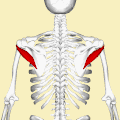
Teres minor muscle (shown in red), seen from behind.
Arm muscles back numbers.png
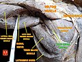
Muscles on the dorsum of the left scapula, and the Triceps brachii muscle:
#3 is Latissimus dorsi muscle
#5 is Teres major muscle
#6 is Teres minor muscle
#7 is Supraspinatus muscle
#8 is Infraspinatus muscle
#13 is long head of Triceps brachii muscle
Details
Origin Lateral border of the scapula
Insertion Inferior facet of greater tubercle of the humerus
Artery Posterior circumflex humeral artery and the circumflex scapular artery
Nerve Axillary nerve (C5-C6)
Actions Laterally rotates the arm, stabilizes humerus
Identifiers
Latin musculus teres minor
TA98 A04.6.02.010
TA2 2459
FMA 32550
Anatomical terms of muscle

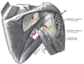
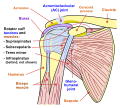
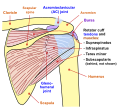
The teres minor (Latin teres meaning 'rounded') is a narrow, elongated muscle of the rotator cuff. The muscle originates from the lateral border and adjacent posterior surface of the corresponding right or left scapula and inserts at both the greater tubercle of the humerus and the posterior surface of the joint capsule. [1]

Contents

The primary function of the teres minor is to modulate the action of the deltoid, preventing the humeral head from sliding upward as the arm is abducted. It also functions to rotate the humerus laterally. The teres minor is innervated by the axillary nerve. [2]

Structure

It arises from the dorsal surface of the axillary border of the scapula for the upper two-thirds of its extent, and from two aponeurotic laminae, one of which separates it from the infraspinatus muscle, the other from the teres major muscle.

Its fibers run obliquely upwards and laterally; the upper ones end in a tendon which is inserted into the lowest of the three impressions on the greater tubercle of the humerus; the lowest fibers are inserted directly into the humerus immediately below this impression.

A normal anatomical variant of this is a much smaller appearing muscle with an intact tendon. This is referred to as the teres minimis; some literature refer to it as teres micro.

Relations

The teres minor originates at the lateral border and adjacent posterior surface of the scapula. It inserts at the greater tubercle of the humerus. The tendon of this muscle passes across, and is united with, the posterior part of the capsule of the shoulder-joint.

Innervation

The muscle is innervated by the posterior branch of axillary nerve where it forms a pseudoganglion. [3] A pseudoganglion has no nerve cells but nerve fibres are present. Damage to the fibers innervating the teres minor is clinically significant.

Variation

Sometimes a group of muscle fibres from teres minor may be fused with infraspinatus.

Function

The infraspinatus and teres minor attach to head of the humerus; as part of the rotator cuff they help hold the humeral head in the glenoid cavity of the scapula. They work in tandem with the posterior deltoid to externally (laterally) rotate the humerus, as well as adduction. Teres Minor can produce only very small scapular plane adduction during maximal contraction (Hughes RE, An KN 1996) with adductor moment arm of approximately 0.2 cm at 45° of shoulder internal rotation and approximately 0.1 cm at 45° of shoulder external rotation.

Clinical significance

Injury

There are two types of rotator cuff injuries: acute tears and chronic tears.

Acute tears occur as a result of a sudden movement. This might include throwing a powerful pitch, holding a fast moving rope during water sports, falling over onto an outstretched hand at speed, or making a sudden thrust with the paddle in kayaking.

A chronic tear develops over a period of time. They usually occur at or near the tendon, as a result of the tendon rubbing against the underlying bone. [4] The teres minor is typically normal following a rotator cuff tear. [5]

Imaging

Conjoint and intact tendon disc of teres minor and infraspinatus muscle. MRI. Conjoint and intact tendon disc of teres minor and infraspinatus muscle..jpg
Conjoint and intact tendon disc of teres minor and infraspinatus muscle.
Intact teres minor tendon with physiologically hypointense signal. MRI. Intact teres minor tendon with physiologically hypointense signal..jpg
Intact teres minor tendon with physiologically hypointense signal.
Hyperintense, fatty degenerated teres minor muscle. MRI. Hyperintense, fatty degenerated teres minor muscle..jpg
Hyperintense, fatty degenerated teres minor muscle.

Atrophy of the teres minor muscle is often a consequence of a rotator cuff tear, but common isolated teres minor atrophies have also been found. A quadrangular space syndrome causes excessive and or chronically compression of the structures which pass through this anatomical tunnel. The axillary nerve and the posterior humeral circumflex artery pass through the space. People affected note shoulder pain and paresthesia down the arm first and foremost in abduction, extension, external rotation and overhead activity. Selective atrophy of the teres minor muscle has been seen and pulled together directly with compression of the corresponding axillary nerve branch or posterior humeral circumflex artery. Fibrous bands, cysts of the glenoid labrum, lipoma or dilated veins can occupy the quadrilateral space pathologically. Similar symptoms are common with anterior shoulder dislocation, humeral neck fracture, brachial plexus injury and thoracic outlet and inlet syndrome. It is important to include those pathologies for a complete as possible differential diagnosis.

Ultrasonography is a tool to detect a fatty degenerative atrophy of the teres minor and shows in affected muscles increased echogenicity and betimes a slight reduction in muscle bulk. MR imaging helps to consolidate the diagnosis of neurogenic muscle atrophy. Extracellular edema after traumatic events causing neural damage show an increased signal intensity on T2-weighted MRI sequences and normal intensity on T1-weighted sequences. Posterior humeral circumflex artery compression and reduced blood flow in stressful arm positions and or maneuvers can be diagnosed by a Doppler ultrasonography. The nerve should be detected adjacent to the vessel. In an elevated arm position the axillary neurovascular bundle can be seen at the posterior axillary fold just before it perforates the deltoideus, while the posterior course is well visible in the neutral position. For a detailed assessment of the artery, a MR angiography is required. The major task of an ultrasonographic examination is to rule out any space occupying mass. Additional electromyography is helpful to reveal any decelerated nerve conduction velocity, and thus denervation of the concerned muscle. [6]

Additional images

See also

Related Research Articles

<span class="mw-page-title-main">Scapula</span> Bone that connects the humerus (upper arm bone) with the clavicle (collar bone)

The scapula, also known as the shoulder blade, is the bone that connects the humerus with the clavicle. Like their connected bones, the scapulae are paired, with each scapula on either side of the body being roughly a mirror image of the other. The name derives from the Classical Latin word for trowel or small shovel, which it was thought to resemble.

<span class="mw-page-title-main">Humerus</span> Long bone of the upper arm

The humerus is a long bone in the arm that runs from the shoulder to the elbow. It connects the scapula and the two bones of the lower arm, the radius and ulna, and consists of three sections. The humeral upper extremity consists of a rounded head, a narrow neck, and two short processes. The body is cylindrical in its upper portion, and more prismatic below. The lower extremity consists of 2 epicondyles, 2 processes, and 3 fossae. As well as its true anatomical neck, the constriction below the greater and lesser tubercles of the humerus is referred to as its surgical neck due to its tendency to fracture, thus often becoming the focus of surgeons.

<span class="mw-page-title-main">Rotator cuff</span> Group of muscles

The rotator cuff is a group of muscles and their tendons that act to stabilize the human shoulder and allow for its extensive range of motion. Of the seven scapulohumeral muscles, four make up the rotator cuff. The four muscles are:

<span class="mw-page-title-main">Axillary nerve</span> Nerve of the human body near the armpit

The axillary nerve or the circumflex nerve is a nerve of the human body, that originates from the brachial plexus at the level of the axilla (armpit) and carries nerve fibers from C5 and C6. The axillary nerve travels through the quadrangular space with the posterior circumflex humeral artery and vein to innervate the deltoid and teres minor.

<span class="mw-page-title-main">Shoulder</span> Part of the body

The human shoulder is made up of three bones: the clavicle (collarbone), the scapula, and the humerus as well as associated muscles, ligaments and tendons.

<span class="mw-page-title-main">Deltoid muscle</span> Shoulder muscle

The deltoid muscle is the muscle forming the rounded contour of the human shoulder. It is also known as the 'common shoulder muscle', particularly in other animals such as the domestic cat. Anatomically, the deltoid muscle is made up of three distinct sets of muscle fibers, namely the

  1. anterior or clavicular part
  2. posterior or scapular part
  3. intermediate or acromial part
<span class="mw-page-title-main">Upper limb</span> Consists of the arm, forearm, and hand

The upper limbs or upper extremities are the forelimbs of an upright-postured tetrapod vertebrate, extending from the scapulae and clavicles down to and including the digits, including all the musculatures and ligaments involved with the shoulder, elbow, wrist and knuckle joints. In humans, each upper limb is divided into the arm, forearm and hand, and is primarily used for climbing, lifting and manipulating objects.

<span class="mw-page-title-main">Triceps</span> Muscle on the back of the upper arm

The triceps, or triceps brachii, is a large muscle on the back of the upper limb of many vertebrates. It consists of 3 parts: the medial, lateral, and long head. It is the muscle principally responsible for extension of the elbow joint.

<span class="mw-page-title-main">Suprascapular nerve</span> Mixed nerve of the upper limb

The suprascapular nerve is a mixed nerve that branches from the upper trunk of the brachial plexus. It is derived from the ventral rami of cervical nerves C5-C6. It provides motor innervation to the supraspinatus muscle, and the infraspinatus muscle.

<span class="mw-page-title-main">Supraspinatus muscle</span> Muscle of the upper back

The supraspinatus is a relatively small muscle of the upper back that runs from the supraspinous fossa superior portion of the scapula to the greater tubercle of the humerus. It is one of the four rotator cuff muscles and also abducts the arm at the shoulder. The spine of the scapula separates the supraspinatus muscle from the infraspinatus muscle, which originates below the spine.

<span class="mw-page-title-main">Axillary artery</span> Large blood vessel bringing oxygenated blood to the thorax

In human anatomy, the axillary artery is a large blood vessel that conveys oxygenated blood to the lateral aspect of the thorax, the axilla (armpit) and the upper limb. Its origin is at the lateral margin of the first rib, before which it is called the subclavian artery.

<span class="mw-page-title-main">Infraspinatus muscle</span> Main external rotator of the shoulder

In human anatomy, the infraspinatus muscle is a thick triangular muscle, which occupies the chief part of the infraspinatous fossa. As one of the four muscles of the rotator cuff, the main function of the infraspinatus is to externally rotate the humerus and stabilize the shoulder joint.

<span class="mw-page-title-main">Shoulder joint</span> Synovial ball and socket joint in the shoulder

The shoulder joint is structurally classified as a synovial ball-and-socket joint and functionally as a diarthrosis and multiaxial joint. It involves an articulation between the glenoid fossa of the scapula and the head of the humerus. Due to the very loose joint capsule ,that gives a limited interface of the humerus and scapula, it is the most mobile joint of the human body.

<span class="mw-page-title-main">Subscapularis muscle</span> Large triangle muscle of the shoulder

The subscapularis is a large triangular muscle which fills the subscapular fossa and inserts into the lesser tubercle of the humerus and the front of the capsule of the shoulder-joint.

<span class="mw-page-title-main">Teres major muscle</span> Muscle of the upper limb

The teres major muscle is a muscle of the upper limb. It attaches to the scapula and the humerus and is one of the seven scapulohumeral muscles. It is a thick but somewhat flattened muscle.

<span class="mw-page-title-main">Dog anatomy</span> Studies of the visible part of a canine

Dog anatomy comprises the anatomical study of the visible parts of the body of a domestic dog. Details of structures vary tremendously from breed to breed, more than in any other animal species, wild or domesticated, as dogs are highly variable in height and weight. The smallest known adult dog was a Yorkshire Terrier that stood only 6.3 cm (2.5 in) at the shoulder, 9.5 cm (3.7 in) in length along the head and body, and weighed only 113 grams (4.0 oz). The heaviest dog was an English Mastiff named Zorba, which weighed 314 pounds (142 kg). The tallest known adult dog is a Great Dane that stands 106.7 cm (42.0 in) at the shoulder.

<span class="mw-page-title-main">Posterior humeral circumflex artery</span>

The posterior humeral circumflex artery arises from the third part of the axillary artery at the distal border of the subscapularis.

<span class="mw-page-title-main">Surgical neck of the humerus</span> Part of one of the bones of the arm

The surgical neck of the humerus is a bony constriction at the proximal end of shaft of humerus. It is situated distal to the greater tubercle and lesser tubercle, and proximal to the deltoid tuberosity.

<span class="mw-page-title-main">Quadrangular space</span>

The quadrangular space, also known as the quadrilateral space (of Velpeau) and the foramen humerotricipitale, is one of the three spaces in the axillary space. The other two spaces are: triangular space and triangular interval.

<span class="mw-page-title-main">Axillary space</span>

The axillary spaces are anatomic spaces. through which axillary contents leave the axilla. They consist of the quadrangular space, triangular space, and triangular interval. It is bounded by teres major, teres minor, medial border of the humerus, and long head of triceps brachii.

References

PD-icon.svgThis article incorporates text in the public domain from page 441 of the 20th edition of Gray's Anatomy (1918)

  1. Saladin, Kenneth (2015). Anatomy & Physiology: The Unity of Form and Function (7 ed.). New York: McGraw-Hill Education. p. 345. ISBN   9780073403717.
  2. Saladin, Kenneth (2015). Anatomy & Physiology: The Unity of Form and Function (7 ed.). New York: McGraw-Hill Education. p. 345. ISBN   9780073403717.
  3. Gitlin, G (October 1957). "Concerning the gangliform enlargement (pseudoganglion) on the nerve to the teres minor muscle". Journal of Anatomy. 91 (4): 466–70. PMC   1244902 . PMID   13475146.
  4. Bahr, Ronald. Ed. Clinical Guide to Sports Injuries. Gazette bok. ISBN   0-7360-4117-6.
  5. Melis, Barbara; DeFranco, Michael; Ladermann, Alexandre; Barthelemy, Renaud; Walch, Gilles (2011). "The teres minor muscle in rotator cuff tendon tears". Skeletal Radiology. 40 (10): 1335–1344. doi:10.1007/s00256-011-1178-3. PMID   21604212. S2CID   8639793 . Retrieved 28 November 2016.
  6. Brestas, P.S.; et al. (September 2006). ". Ultrasound findings of teres minor denervation in suspected quadrilateral space syndrome". J Clin Ultrasound. 34 (7): 343–7. doi:10.1002/jcu.20239. PMID   16869012. S2CID   6341877.