 The palmar aponeurosis
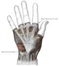
| Palmaris brevis muscle | |
|---|---|
 The muscles of the left hand. Palmar surface (palmaris brevis visible at center left). | |
| Details | |
| Origin | Flexor retinaculum (medial) and palmar aponeurosis |
| Insertion | Palm |
| Artery | Palmar metacarpal artery |
| Nerve | Superficial branch of ulnar nerve |
| Actions | Pulls on skin over hypothenar eminence, deepening the cup of the palm and so improving grip |
| Identifiers | |
| Latin | musculus palmaris brevis |
| TA98 | A04.6.02.053 |
| TA2 | 2520 |
| FMA | 37381 |
| Anatomical terms of muscle | |
Palmaris brevis muscle is a thin, quadrilateral muscle, placed beneath the integument of the ulnar side of the hand. It acts to fold the skin of the hypothenar eminence transversally.
