Brachialis | |
---|---|
Details | |
Origin | Anterior surface of the humerus, particularly the distal half of this bone |
Insertion | Coronoid process and the tuberosity of the ulna |
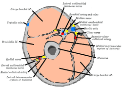
Artery | Radial recurrent artery, brachial artery |
Nerve | Musculocutaneous nerve (C5-C7) and radial nerve (C5, C6) |
Actions | Flexion at elbow joint |
Identifiers | |
Latin | musculus brachialis |
TA98 | A04.6.02.018 |
TA2 | 2469 |
FMA | 37667 |
Anatomical terms of muscle |
The brachialis (brachialis anticus) is a muscle in the upper arm that flexes the elbow. It lies beneath the biceps brachii, and makes up part of the floor of the region known as the cubital fossa (elbow pit). It originates from the anterior aspect of the distal humerus; [1] it inserts onto the tuberosity of the ulna. It is innervated by the musculocutaneous nerve, [2] and commonly also receives additional innervation from the radial nerve. [3] The brachialis is the prime mover of elbow flexion generating about 50% more power than the biceps.[ dubious ] [1]
The brachialis originates from the anterior surface of the distal half of the humerus, [1] near the insertion of the deltoid muscle, which it embraces by two angular processes. Its origin extends below to within 2.5 cm of the margin of the articular surface of the humerus at the elbow joint. [2]
Its fibers converge to a thick tendon which is inserted into the tuberosity of the ulna, [2] and the rough depression on the anterior surface of the coronoid process of the ulna.[ citation needed ]
The brachialis muscle is innervated by the musculocutaneous nerve, which runs on its superficial surface, between it and the biceps brachii. [2] However, in 70-80% of people, the muscle has double innervation with the radial nerve (C5-T1). The divide between the two innervations is at the insertion of the deltoid. [3]
The brachialis is supplied by muscular branches of the brachial artery and by the recurrent radial artery. [4]
The muscle is occasionally doubled; additional muscle slips to the supinator, pronator teres, biceps brachii, lacertus fibrosus, or radius are more rarely found.[ citation needed ]
The brachialis flexes the arm at the elbow joint. [2] Unlike the biceps, the brachialis does not insert on the radius, and does not participate in pronation and supination of the forearm. [2]
The brachialis muscle [5] In classical Latin bracchialis means of or belonging to the arm, [6] and is derived from classical Latin bracchium, "arm". [6] The expression musculus brachialis is used in the current official anatomic nomenco Terminologia Anatomica . [7]
In human anatomy, the arm refers to the upper limb in common usage, although academically the term specifically means the upper arm between the glenohumeral joint and the elbow joint. The distal part of the upper limb between the elbow and the radiocarpal joint is known as the forearm or "lower" arm, and the extremity beyond the wrist is the hand.
The ulna or ulnar bone is a long bone in the forearm stretching from the elbow to the wrist. It is on the same side of the forearm as the little finger, running parallel to the radius, the forearm's other long bone. Longer and thinner than the radius, the ulna is considered to be the smaller long bone of the lower arm. The corresponding bone in the lower leg is the fibula.
The humerus is a long bone in the arm that runs from the shoulder to the elbow. It connects the scapula and the two bones of the lower arm, the radius and ulna, and consists of three sections. The humeral upper extremity consists of a rounded head, a narrow neck, and two short processes. The body is cylindrical in its upper portion, and more prismatic below. The lower extremity consists of 2 epicondyles, 2 processes, and 3 fossae. As well as its true anatomical neck, the constriction below the greater and lesser tubercles of the humerus is referred to as its surgical neck due to its tendency to fracture, thus often becoming the focus of surgeons.
The biceps or biceps brachii are a large muscle that lies on the front of the upper arm between the shoulder and the elbow. Both heads of the muscle arise on the scapula and join to form a single muscle belly which is attached to the upper forearm. While the biceps crosses both the shoulder and elbow joints, its main function is at the elbow where it flexes the forearm and supinates the forearm. Both these movements are used when opening a bottle with a corkscrew: first biceps screws in the cork (supination), then it pulls the cork out (flexion).
The brachioradialis is a muscle of the forearm that flexes the forearm at the elbow. It is also capable of both pronation and supination, depending on the position of the forearm. It is attached to the distal styloid process of the radius by way of the brachioradialis tendon, and to the lateral supracondylar ridge of the humerus.
The radial nerve is a nerve in the human body that supplies the posterior portion of the upper limb. It innervates the medial and lateral heads of the triceps brachii muscle of the arm, as well as all 12 muscles in the posterior osteofascial compartment of the forearm and the associated joints and overlying skin.
The upper limbs or upper extremities are the forelimbs of an upright-postured tetrapod vertebrate, extending from the scapulae and clavicles down to and including the digits, including all the musculatures and ligaments involved with the shoulder, elbow, wrist and knuckle joints. In humans, each upper limb is divided into the arm, forearm and hand, and is primarily used for climbing, lifting and manipulating objects.
The musculocutaneous nerve is a mixed branch of the lateral cord of the brachial plexus derived from cervical spinal nerves C5-C7. It arises opposite the lower border of the pectoralis major. It provides motor innervation to the muscles of the anterior compartment of the arm: the coracobrachialis, biceps brachii, and brachialis. It provides sensory innervation to the lateral forearm. It courses through the anterior part of the arm, terminating 2 cm above elbow; after passing the lateral edge of the tendon of biceps brachii it is becomes known as the lateral cutaneous nerve of the forearm.
The triceps, or triceps brachii, is a large muscle on the back of the upper limb of many vertebrates. It consists of 3 parts: the medial, lateral, and long head. It is the muscle principally responsible for extension of the elbow joint.
The cubital fossa, chelidon or inside of elbow is the area on the anterior side of the upper part between the arm and forearm of a human or other hormid animals. It lies anteriorly to the elbow when in standard anatomical position.
The anconeus muscle is a small muscle on the posterior aspect of the elbow joint.
The coracobrachialis muscle is a muscle in the upper medial part of the arm. It is located within the anterior compartment of the arm. It originates from the coracoid process of the scapula; it inserts onto the middle of the medial aspect of the body of the humerus. It is innervated by the musculocutaneous nerve. It acts to adduct and flex the arm.
The pronator teres is a muscle that, along with the pronator quadratus, serves to pronate the forearm. It has two origins, at the medial humeral supracondylar ridge and the ulnar tuberosity, and inserts near the middle of the radius.
In human anatomy, the supinator is a broad muscle in the posterior compartment of the forearm, curved around the upper third of the radius. Its function is to supinate the forearm.
Dog anatomy comprises the anatomical study of the visible parts of the body of a domestic dog. Details of structures vary tremendously from breed to breed, more than in any other animal species, wild or domesticated, as dogs are highly variable in height and weight. The smallest known adult dog was a Yorkshire Terrier that stood only 6.3 cm (2.5 in) at the shoulder, 9.5 cm (3.7 in) in length along the head and body, and weighed only 113 grams (4.0 oz). The heaviest dog was an English Mastiff named Zorba, which weighed 314 pounds (142 kg). The tallest known adult dog is a Great Dane that stands 106.7 cm (42.0 in) at the shoulder.
The deep artery of arm is a large artery of the arm which arises from the brachial artery. It descends in the arm before ending by anastomosing with the radial recurrent artery.
The fascial compartments of arm refers to the specific anatomical term of the compartments within the upper segment of the upper limb of the body. The upper limb is divided into two segments, the arm and the forearm. Each of these segments is further divided into two compartments which are formed by deep fascia – tough connective tissue septa (walls). Each compartment encloses specific muscles and nerves.
The coronoid process of the ulna is a triangular process projecting forward from the anterior proximal portion of the ulna.
The humeroulnar joint is part of the elbow-joint. It is composed of two bones, the humerus and ulna, and is the junction between the trochlear notch of ulna and the trochlea of humerus. It is classified as a simple hinge-joint, which allows for movements of flexion, extension and circumduction. Owing to the obliquity of the trochlea of the humerus, this movement does not take place in the antero-posterior plane of the body of the humerus.
The elbow is the region between the upper arm and the forearm that surrounds the elbow joint. The elbow includes prominent landmarks such as the olecranon, the cubital fossa, and the lateral and the medial epicondyles of the humerus. The elbow joint is a hinge joint between the arm and the forearm; more specifically between the humerus in the upper arm and the radius and ulna in the forearm which allows the forearm and hand to be moved towards and away from the body. The term elbow is specifically used for humans and other primates, and in other vertebrates it is not used. In those cases, forelimb plus joint is used.
This article incorporates text in the public domain from page 444 of the 20th edition of Gray's Anatomy (1918)