The carpal bones are the eight small bones that make up the wrist (carpus) that connects the hand to the forearm. The term "carpus" and "carpal" is derived from the Latin carpus and the Greek καρπός (karpós), meaning "wrist". In human anatomy, the main role of the carpal bones is to articulate with the radial and ulnar heads to form a highly mobile condyloid joint, to provide attachments for thenar and hypothenar muscles, and to form part of the rigid carpal tunnel which allows the median nerve and tendons of the anterior forearm muscles to be transmitted to the hand and fingers.

The median nerve is a nerve in humans and other animals in the upper limb. It is one of the five main nerves originating from the brachial plexus.

In human anatomy, the wrist is variously defined as (1) the carpus or carpal bones, the complex of eight bones forming the proximal skeletal segment of the hand; (2) the wrist joint or radiocarpal joint, the joint between the radius and the carpus and; (3) the anatomical region surrounding the carpus including the distal parts of the bones of the forearm and the proximal parts of the metacarpus or five metacarpal bones and the series of joints between these bones, thus referred to as wrist joints. This region also includes the carpal tunnel, the anatomical snuff box, bracelet lines, the flexor retinaculum, and the extensor retinaculum.
The flexor digitorum profundus is a muscle in the forearm of humans that flexes the fingers. It is considered an extrinsic hand muscle because it acts on the hand while its muscle belly is located in the forearm.

In human anatomy, the ulnar nerve is a nerve that runs near the ulna bone. The ulnar collateral ligament of elbow joint is in relation with the ulnar nerve. The nerve is the largest in the human body unprotected by muscle or bone, so injury is common. This nerve is directly connected to the little finger, and the adjacent half of the ring finger, innervating the palmar aspect of these fingers, including both front and back of the tips, perhaps as far back as the fingernail beds.

In human anatomy, the metacarpal bones or metacarpus, also known as the "palm bones", are the appendicular bones that form the intermediate part of the hand between the phalanges (fingers) and the carpal bones, which articulate with the forearm. The metacarpal bones are homologous to the metatarsal bones in the foot.

The hamate bone, or unciform bone, Latin os hamatum and occasionally abbreviated as just hamatum, is a bone in the human wrist readily distinguishable by its wedge shape and a hook-like process ("hamulus") projecting from its palmar surface.

The pisiform bone, also spelled pisiforme, is a small knobbly, sesamoid bone that is found in the wrist. It forms the ulnar border of the carpal tunnel.

The ulnar artery is the main blood vessel, with oxygenated blood, of the medial aspects of the forearm. It arises from the brachial artery and terminates in the superficial palmar arch, which joins with the superficial branch of the radial artery. It is palpable on the anterior and medial aspect of the wrist.

The flexor pollicis brevis is a muscle in the hand that flexes the thumb. It is one of three thenar muscles. It has both a superficial part and a deep part.
The pronator teres is a muscle that, along with the pronator quadratus, serves to pronate the forearm. It has two origins, at the medial humeral supracondylar ridge and the ulnar tuberosity, and inserts near the middle of the radius.

The palmaris longus is a muscle visible as a small tendon located between the flexor carpi radialis and the flexor carpi ulnaris, although it is not always present. It is absent in about 14 percent of the population; this number can vary in African, Asian, and Native American populations, however. Absence of the palmaris longus does not have an effect on grip strength. The lack of palmaris longus muscle does result in decreased pinch strength in fourth and fifth fingers. The absence of palmaris longus muscle is more prevalent in females than males.

In human anatomy, the abductor digiti minimi is a skeletal muscle situated on the ulnar border of the palm of the hand. It forms the ulnar border of the palm and its spindle-like shape defines the hypothenar eminence of the palm together with the skin, connective tissue, and fat surrounding it. Its main function is to pull the little finger away from the other fingers.
The palmar carpal ligament is a thickened portion of antebrachial fascia on anterior/palmar side of the wrist which - together with the flexor retinaculum of the hand - retains the tendons of most of the flexor muscles of the hand.

The ulnar canal or ulnar tunnel (also known as Guyon's canal or tunnel) is a semi-rigid longitudinal canal in the wrist that allows passage of the ulnar artery and ulnar nerve into the hand. The roof of the canal is made up of the superficial palmar carpal ligament, while the deeper flexor retinaculum and hypothenar muscles comprise the floor. The space is medially bounded by the pisiform and pisohamate ligament more proximally, and laterally bounded by the hook of the hamate more distally. It is approximately 4 cm long, beginning proximally at the transverse carpal ligament and ending at the aponeurotic arch of the hypothenar muscles.

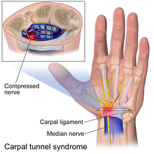
In the human body, the carpal tunnel or carpal canal is a flattened body cavity on the flexor (palmar/volar) side of the wrist, bounded by the carpal bones and flexor retinaculum. It forms the passageway that transmits the median nerve and the tendons of the extrinsic flexor muscles of the hand from the forearm to the hand. There are described cases of the anatomical variant median artery occurrence.

A hand is a prehensile, multi-fingered appendage located at the end of the forearm or forelimb of primates such as humans, chimpanzees, monkeys, and lemurs. A few other vertebrates such as the koala are often described as having "hands" instead of paws on their front limbs. The raccoon is usually described as having "hands" though opposable thumbs are lacking.

Wrist pain or open wrist is a syndrome inhibiting use of a hand due to pain in anatomical structures of the wrist. It most commonly results from an injury to a ligament. The pain may be sharp from a traumatic injury or from chronic repetitive wrist activities.
Injuries to the arm, forearm or wrist area can lead to various nerve disorders. One such disorder is median nerve palsy. The median nerve controls the majority of the muscles in the forearm. It controls abduction of the thumb, flexion of hand at wrist, flexion of digital phalanx of the fingers, is the sensory nerve for the first three fingers, etc. Because of this major role of the median nerve, it is also called the eye of the hand. If the median nerve is damaged, the ability to abduct and oppose the thumb may be lost due to paralysis of the thenar muscles. Various other symptoms can occur which may be repaired through surgery and tendon transfers. Tendon transfers have been very successful in restoring motor function and improving functional outcomes in patients with median nerve palsy.

The muscles of the thumb are nine skeletal muscles located in the hand and forearm. The muscles allow for flexion, extension, adduction, abduction and opposition of the thumb. The muscles acting on the thumb can be divided into two groups: The extrinsic hand muscles, with their muscle bellies located in the forearm, and the intrinsic hand muscles, with their muscles bellies located in the hand proper.