| Flexor digitorum superficialis | |
|---|---|
 | |
 Flexor digitorum superficialis | |
| Details | |
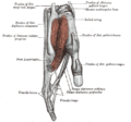
| Origin | Medial epicondyle of the humerus (common flexor tendon) as well as parts of the radius and ulna |
| Insertion | Anterior margins on the base of the middle phalanges of the four fingers |
| Artery | Radial artery |
| Nerve | Median nerve |
| Actions | Flexor of fingers (primarily at proximal interphalangeal joints) |
| Antagonist | Extensor digitorum muscle |
| Identifiers | |
| Latin | musculus flexor digitorum superficialis |
| TA98 | A04.6.02.033 |
| TA2 | 2486 |
| FMA | 38469 |
| Anatomical terms of muscle | |
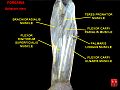
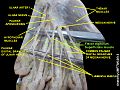
Flexor digitorum superficialis (flexor digitorum sublimis) or flexor digitorum communis sublimis [1] is an extrinsic flexor muscle of the fingers at the proximal interphalangeal joints.
Contents
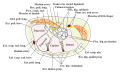
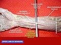
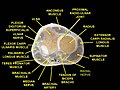
It is in the anterior compartment of the forearm. It is sometimes considered to be the deepest part of the superficial layer of this compartment, [2] [3] and sometimes considered to be a distinct, "intermediate layer" of this compartment. [4] It is relatively common for the flexor digitorum superficialis to be missing from the little finger, bilaterally and unilaterally, which can cause problems when diagnosing a little finger injury. [5]