Adductor pollicis muscle

Last updated
Adductor pollicis muscle
1121 Intrinsic Muscles of the Hand Superficial sin.png
The superficial muscles of the left hand. Palmar view
Adductor pollicis is labelled at bottom right.
Details
Origin Transverse head: anterior body of the third metacarpal
Oblique head: bases of the second and the third metacarpals and the adjacent trapezoid and capitate bones
Insertion medial side of the base of the proximal phalanx of the thumb and the ulnar sesamoid
Artery deep palmar arch
Nerve deep branch of the ulnar nerve (T1)
Actions adducts the thumb at the carpometacarpal joint
Antagonist Abductor pollicis longus muscle, Abductor pollicis brevis muscle
Identifiers
Latin musculus adductor pollicis
TA98 A04.6.02.059
TA2 2526
FMA 37380
Anatomical terms of muscle

In human anatomy, the adductor pollicis muscle is a muscle in the hand that functions to adduct the thumb. It has two heads: transverse and oblique.

Contents

It is a fleshy, flat, triangular, and fan-shaped muscle deep in the thenar compartment beneath the long flexor tendons and the lumbrical muscles at the center of the palm. It overlies the metacarpal bones and the interosseous muscles. [1]

Structure

Oblique head

The oblique head (Latin: adductor obliquus pollicis) arises by several slips from the capitate bone, the bases of the second and third metacarpals, the intercarpal ligaments, and the sheath of the tendon of the flexor carpi radialis. [2]

From this origin the greater number of fibers pass obliquely downward and converge to a tendon, which, uniting with the tendons of the medial portion of the flexor pollicis brevis and the transverse head of the adductor pollicis, is inserted into the ulnar side of the base of the proximal phalanx of the thumb, a sesamoid bone being present in the tendon. [2]

A considerable fasciculus, however, passes more obliquely beneath the tendon of the flexor pollicis longus to join the lateral portion of the flexor pollicis brevis and the abductor pollicis brevis. [2]

Transverse head

The transverse head (Latin: adductor transversus pollicis) is deeply seated. [2]

It is triangular, arising by a broad base from the lower two-thirds of the palmar surface of the third metacarpal bone; the fibers converge, to be inserted with the medial part of the flexor pollicis brevis and the oblique head into the ulnar side of the base of the proximal phalanx of the thumb. [2]

Relations

The radial artery passes between the two heads, travelling from the back of the hand into the palm, where it forms the deep palmar arch.

Innervation

The adductor pollicis is innervated by the deep branch of the ulnar nerve (C8–T1). [3]

Between the oblique and transverse heads is a thin fibrous arcade which the nerve passes as it traverses the palm laterally. The nerve is accompanied by the deep palmar arch. [1]

Function

While adduction of the thumb (bringing it back into the plane of the palm of the hand from its previously abducted position) is mainly produced by the adductor pollicis, it can also bring the thumb to the side of the palm and index finger and the flexor pollicis brevis and the opponens pollicis help in thumb adduction. [3]

Clinical significance

Froment's sign is used to test for a compromised adductor pollicis muscle.

In neuromuscular monitoring, the ulnar nerve is stimulated and the strength of adductor pollicis contraction is measured.

Other animals

The adductor pollicis evolved from the contrahens I muscle as man's ancestors' thumbs and big toes became opposable. It might also contain an element of the thumb's interosseous muscle. [4]

In the Pan-Homo LCA the oblique head of the adductor pollicis probably had a relatively small physiological cross sectional area (PCSA) and both heads probably acted as extensors and adductors at the carpometacarpal joint. In humans the PCSA of the oblique head is relatively enlarged and both heads act as flexors at this joint. [5]

See also

Additional images

Related Research Articles

<span class="mw-page-title-main">Carpal bones</span> Eight small bones that make up the wrist (or carpus) that connects the hand to the forearm

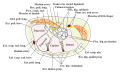
The carpal bones are the eight small bones that make up the wrist (carpus) that connects the hand to the forearm. The term "carpus" and "carpal" is derived from the Latin carpus and the Greek καρπός (karpós), meaning "wrist". In human anatomy, the main role of the carpal bones is to articulate with the radial and ulnar heads to form a highly mobile condyloid joint, to provide attachments for thenar and hypothenar muscles, and to form part of the rigid carpal tunnel which allows the median nerve and tendons of the anterior forearm muscles to be transmitted to the hand and fingers.

<span class="mw-page-title-main">Thumb</span> First digit of the hand

The thumb is the first digit of the hand, next to the index finger. When a person is standing in the medical anatomical position, the thumb is the outermost digit. The Medical Latin English noun for thumb is pollex, and the corresponding adjective for thumb is pollical.

<span class="mw-page-title-main">Wrist</span> Part of the arm between the lower arm and the hand

In human anatomy, the wrist is variously defined as (1) the carpus or carpal bones, the complex of eight bones forming the proximal skeletal segment of the hand; (2) the wrist joint or radiocarpal joint, the joint between the radius and the carpus and; (3) the anatomical region surrounding the carpus including the distal parts of the bones of the forearm and the proximal parts of the metacarpus or five metacarpal bones and the series of joints between these bones, thus referred to as wrist joints. This region also includes the carpal tunnel, the anatomical snuff box, bracelet lines, the flexor retinaculum, and the extensor retinaculum.

<span class="mw-page-title-main">Thenar eminence</span> Mound at the base of the thumb formed by the intrinsic group of muscles

The thenar eminence is the mound formed at the base of the thumb on the palm of the hand by the intrinsic group of muscles of the thumb. The skin overlying this region is the area stimulated when trying to elicit a palmomental reflex. The word thenar comes from Ancient Greek θέναρ (thenar) 'palm of the hand'.

<span class="mw-page-title-main">Trapezium (bone)</span> Bone of the wrist

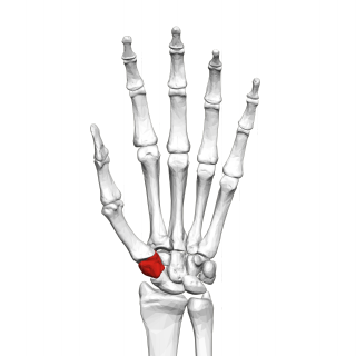
The trapezium bone is a carpal bone in the hand. It forms the radial border of the carpal tunnel.

<span class="mw-page-title-main">Upper limb</span> Consists of the arm, forearm, and hand

The upper limbs or upper extremities are the forelimbs of an upright-postured tetrapod vertebrate, extending from the scapulae and clavicles down to and including the digits, including all the musculatures and ligaments involved with the shoulder, elbow, wrist and knuckle joints. In humans, each upper limb is divided into the arm, forearm and hand, and is primarily used for climbing, lifting and manipulating objects.

In human anatomy, the extensor pollicis longus muscle (EPL) is a skeletal muscle located dorsally on the forearm. It is much larger than the extensor pollicis brevis, the origin of which it partly covers and acts to stretch the thumb together with this muscle.

<span class="mw-page-title-main">Flexor pollicis brevis muscle</span> Muscle in the thenar compartment

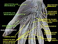
The flexor pollicis brevis is a muscle in the hand that flexes the thumb. It is one of three thenar muscles. It has both a superficial part and a deep part.

The flexor pollicis longus is a muscle in the forearm and hand that flexes the thumb. It lies in the same plane as the flexor digitorum profundus. This muscle is unique to humans, being either rudimentary or absent in other primates. A meta-analysis indicated accessory flexor pollicis longus is present in around 48% of the population.

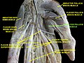
In human anatomy, the abductor pollicis longus (APL) is one of the extrinsic muscles of the hand. Its major function is to abduct the thumb at the wrist. Its tendon forms the anterior border of the anatomical snuffbox.

<span class="mw-page-title-main">Palmar interossei muscles</span> Muscles between the metacarpals

In human anatomy, the palmar or volar interossei are four muscles, one on the thumb that is occasionally missing, and three small, unipennate, central muscles in the hand that lie between the metacarpal bones and are attached to the index, ring, and little fingers. They are smaller than the dorsal interossei of the hand.

<span class="mw-page-title-main">Carpometacarpal joint</span>

The carpometacarpal (CMC) joints are five joints in the wrist that articulate the distal row of carpal bones and the proximal bases of the five metacarpal bones.

<span class="mw-page-title-main">Dorsal interossei of the hand</span> Muscles between the metacarpals

In human anatomy, the dorsal interossei (DI) are four muscles in the back of the hand that act to abduct (spread) the index, middle, and ring fingers away from hand's midline and assist in flexion at the metacarpophalangeal joints and extension at the interphalangeal joints of the index, middle and ring fingers.

<span class="mw-page-title-main">Palmar aponeurosis</span>

The palmar aponeurosis invests the muscles of the palm, and consists of central, lateral, and medial portions.

<span class="mw-page-title-main">Deep palmar arch</span>

The deep palmar arch is an arterial network found in the palm. It is usually primarily formed from the terminal part of the radial artery. The ulnar artery also contributes through an anastomosis. This is in contrast to the superficial palmar arch, which is formed predominantly by the ulnar artery.

<span class="mw-page-title-main">Princeps pollicis artery</span> Artery of the human thumb

The princeps pollicis artery, or principal artery of the thumb, arises from the radial artery just as it turns medially towards the deep part of the hand; it descends between the first dorsal interosseous muscle and the oblique head of the adductor pollicis, along the medial side of the first metacarpal bone to the base of the proximal phalanx, where it lies beneath the tendon of the flexor pollicis longus muscle and divides into two branches.

<span class="mw-page-title-main">Deep branch of ulnar nerve</span>

The deep branch of the ulnar nerve is a terminal, primarily motor branch of the ulnar nerve. It is accompanied by the deep palmar branch of ulnar artery.

<span class="mw-page-title-main">Outline of human anatomy</span> Overview of and topical guide to human anatomy

The following outline is provided as an overview of and topical guide to human anatomy:

<span class="mw-page-title-main">Extrinsic extensor muscles of the hand</span>

The extrinsic extensor muscles of the hand are located in the back of the forearm and have long tendons connecting them to bones in the hand, where they exert their action. Extrinsic denotes their location outside the hand. Extensor denotes their action which is to extend, or open flat, joints in the hand. They include the extensor carpi radialis longus (ECRL), extensor carpi radialis brevis (ECRB), extensor digitorum (ED), extensor digiti minimi (EDM), extensor carpi ulnaris (ECU), abductor pollicis longus (APL), extensor pollicis brevis (EPB), extensor pollicis longus (EPL), and extensor indicis (EI).

<span class="mw-page-title-main">Muscles of the thumb</span>

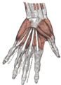
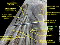
The muscles of the thumb are nine skeletal muscles located in the hand and forearm. The muscles allow for flexion, extension, adduction, abduction and opposition of the thumb. The muscles acting on the thumb can be divided into two groups: The extrinsic hand muscles, with their muscle bellies located in the forearm, and the intrinsic hand muscles, with their muscles bellies located in the hand proper.

References

PD-icon.svgThis article incorporates text in the public domain from page 462 of the 20th edition of Gray's Anatomy (1918)

  1. 1 2 Chang & Blair 1985
  2. 1 2 3 4 5 Gray's Anatomy 1918. (See infobox)
  3. 1 2 Platzer 2004 , p. 176
  4. Yamamoto, Murakami & Ohtsuka 1988
  5. Tocheri et al. 2008 , p. 549