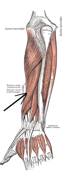
Abductor pollicis longus muscle | |
---|---|
Details | |
Origin | Ulna, radius, Interosseous membrane |
Insertion | Base of 1st metacarpal |
Artery | Posterior interosseous artery |
Nerve | Radial nerve C8, T1 |
Actions | Abduction, extension of thumb |
Antagonist | Adductor pollicis muscle |
Identifiers | |
Latin | musculus abductor pollicis longus |
TA98 | A04.6.02.049 |
TA2 | 2517 |
FMA | 38515 |
Anatomical terms of muscle |
In human anatomy, the abductor pollicis longus (APL) is one of the extrinsic muscles of the hand. Its major function is to abduct the thumb at the wrist. Its tendon forms the anterior border of the anatomical snuffbox.
The abductor pollicis longus lies immediately below the supinator and is sometimes united with it. It arises from the lateral part of the dorsal surface of the body of the ulna, [1] below the insertion of the anconeus, from the interosseous membrane, and from the middle third of the dorsal surface of the body of the radius. [2]
Passing obliquely downward and lateralward, it ends in a tendon, which runs through a groove on the lateral side of the lower end of the radius, accompanied by the tendon of the extensor pollicis brevis. [2]
The insertion is divided into a distal, superficial part and a proximal, deep part. The superficial part is inserted with one or more tendons into the radial side of the base of the first metacarpal bone, and the deep part is variably inserted into the trapezium, the joint capsule and its ligaments, and into the belly of abductor pollicis brevis (APB) or opponens pollicis. [3]
The abductor pollicis longus muscle is innervated by the posterior interosseous nerve, which is a continuation of the deep branch of the radial nerve after it passes through the supinator muscle. Abductor pollicis longus lies close to the radial nerve. [4] The posterior interosseous nerve is derived from spinal segments C7 & C8. [5]
Abductor pollicis longus is supplied by the posterior interosseous artery. [6]
An accessory abductor pollicis longus (AAPL) tendon is present in more than 80% of people, and a separate muscle belly is present in 20% of people. In one study, the accessory tendon was inserted into the trapezium (41%); proximally on the abductor pollicis brevis (22%) and opponens pollicis brevis (5%); had a double insertion on the trapezium and thenar muscles (15%); or the base of the first metacarpal (1%). [7] Up to seven tendons have been reported in rare cases. [8]
Multiple APL tendons can be regarded as a functional advantage since injured tendons can be compensated by the healthy ones. [9]
The chief action of abductor pollicis longus is to abduct the thumb at the carpometacarpal joint, thereby moving the thumb anteriorly. It also assists in extending and rotating the thumb. [6]
By its continued action, it helps to abduct the wrist (radial deviation) and flex the hand. [6]
The APL insertion on the trapezium and the APB origin on the same bone is the only connection between the thumb's intrinsic and extrinsic muscles. [lower-alpha 1] As the thumb is brought into action, these two muscles must coordinate to keep the trapezium stable in the carpus, [3] which is important for the proper functioning of the thumb (i.e. precision and power grip.) [10]
The only primates to have an APL completely separated from the extensor pollicis brevis are modern humans and gibbons. [11] In gibbons, however, the APL originates proximally on the radius and ulna, whereas it originates in the middle part of these bones in crab-eating monkeys, bonobos, and humans. In all these primates, the muscle is inserted onto the base of the first metacarpal and sometimes onto the trapezium (siamangs and bonobos) and thumb sesamoids (crab-eating monkeys). [12]
In chimpanzees, the APL flexes the thumb rather than extending it like in modern humans. Compared to the wrists of chimpanzees, the human wrist is derived (compared to the Pan-Homo LCA) in having considerably longer muscle moment arms for a range of hand muscles. It is possible that these differences are due to the supinated position of the trapezium in humans which, in its turn, is a result of the expansion of the trapezoid on the side of the palm. [13]
A small, lens-shaped radial sesamoid embedded into the APL tendon is a primitive state found in all known Carnivora genera except in the red and giant pandas and the extinct Simocyon where it is hypertrophied (enlarged) into a sixth digit or a so-called "false thumb", a derived trait that first appeared in ursids. [14] The APL sesamoid is present in all non-human primates, but only in about half of gorillas, and normally absent in humans. [15]
A finger is a prominent digit on the forelimbs of most tetrapod vertebrate animals, especially those with prehensile extremities such as humans and other primates. Most tetrapods have five digits (pentadactyly), and short digits are typically referred to as toes, while those that are notably elongated are called fingers. In humans, the fingers are flexibly articulated and opposable, serving as an important organ of tactile sensation and fine movements, which are crucial to the dexterity of the hands and the ability to grasp and manipulate objects.
The thumb is the first digit of the hand, next to the index finger. When a person is standing in the medical anatomical position, the thumb is the outermost digit. The Medical Latin English noun for thumb is pollex, and the corresponding adjective for thumb is pollical.
The radial nerve is a nerve in the human body that supplies the posterior portion of the upper limb. It innervates the medial and lateral heads of the triceps brachii muscle of the arm, as well as all 12 muscles in the posterior osteofascial compartment of the forearm and the associated joints and overlying skin.
The anatomical snuff box or snuffbox or foveola radialis is a triangular deepening on the radial, dorsal aspect of the hand—at the level of the carpal bones, specifically, the scaphoid and trapezium bones forming the floor. The name originates from the use of this surface for placing and then sniffing powdered tobacco, or "snuff." It is sometimes referred to by its French name tabatière.
The extensor carpi radialis longus is one of the five main muscles that control movements at the wrist. This muscle is quite long, starting on the lateral side of the humerus, and attaching to the base of the second metacarpal bone.
In human anatomy, the metacarpal bones or metacarpus, also known as the "palm bones", are the appendicular bones that form the intermediate part of the hand between the phalanges (fingers) and the carpal bones, which articulate with the forearm. The metacarpal bones are homologous to the metatarsal bones in the foot.
Wrist drop is a medical condition in which the wrist and the fingers cannot extend at the metacarpophalangeal joints. The wrist remains partially flexed due to an opposing action of flexor muscles of the forearm. As a result, the extensor muscles in the posterior compartment remain paralyzed.
The upper limbs or upper extremities are the forelimbs of an upright-postured tetrapod vertebrate, extending from the scapulae and clavicles down to and including the digits, including all the musculatures and ligaments involved with the shoulder, elbow, wrist and knuckle joints. In humans, each upper limb is divided into the arm, forearm and hand, and is primarily used for climbing, lifting and manipulating objects.
In human anatomy, the extensor pollicis longus muscle (EPL) is a skeletal muscle located dorsally on the forearm. It is much larger than the extensor pollicis brevis, the origin of which it partly covers and acts to stretch the thumb together with this muscle.
The flexor pollicis longus is a muscle in the forearm and hand that flexes the thumb. It lies in the same plane as the flexor digitorum profundus. This muscle is unique to humans, being either rudimentary or absent in other primates. A meta-analysis indicated accessory flexor pollicis longus is present in around 48% of the population.
In human anatomy, the adductor pollicis muscle is a muscle in the hand that functions to adduct the thumb. It has two heads: transverse and oblique.
In human anatomy, the extensor pollicis brevis (EPB) is a skeletal muscle on the dorsal side of the forearm. It lies on the medial side of, and is closely connected with, the abductor pollicis longus. The extensor pollicis brevis belongs to the deep group of the posterior fascial compartment of the forearm. It is a part of the lateral border of the anatomical snuffbox.
In human anatomy, the palmar or volar interossei are four muscles, one on the thumb that is occasionally missing, and three small, unipennate, central muscles in the hand that lie between the metacarpal bones and are attached to the index, ring, and little fingers. They are smaller than the dorsal interossei of the hand.
The carpometacarpal (CMC) joints are five joints in the wrist that articulate the distal row of carpal bones and the proximal bases of the five metacarpal bones.
In human anatomy, the abductor digiti minimi is a skeletal muscle situated on the ulnar border of the palm of the hand. It forms the ulnar border of the palm and its spindle-like shape defines the hypothenar eminence of the palm together with the skin, connective tissue, and fat surrounding it. Its main function is to pull the little finger away from the other fingers.
The posterior compartment of the forearm contains twelve muscles which primarily extend the wrist and digits. It is separated from the anterior compartment by the interosseous membrane between the radius and ulna.
The cervical spinal nerve 8 (C8) is a spinal nerve of the cervical segment.
The muscles of the hand are the skeletal muscles responsible for the movement of the hand and fingers. The muscles of the hand can be subdivided into two groups: the extrinsic and intrinsic muscle groups. The extrinsic muscle groups are the long flexors and extensors. They are called extrinsic because the muscle belly is located on the forearm. The intrinsic group are the smaller muscles located within the hand itself. The muscles of the hand are innervated by the radial, median, and ulnar nerves from the brachial plexus.
The extrinsic extensor muscles of the hand are located in the back of the forearm and have long tendons connecting them to bones in the hand, where they exert their action. Extrinsic denotes their location outside the hand. Extensor denotes their action which is to extend, or open flat, joints in the hand. They include the extensor carpi radialis longus (ECRL), extensor carpi radialis brevis (ECRB), extensor digitorum (ED), extensor digiti minimi (EDM), extensor carpi ulnaris (ECU), abductor pollicis longus (APL), extensor pollicis brevis (EPB), extensor pollicis longus (EPL), and extensor indicis (EI).
The muscles of the thumb are nine skeletal muscles located in the hand and forearm. The muscles allow for flexion, extension, adduction, abduction and opposition of the thumb. The muscles acting on the thumb can be divided into two groups: The extrinsic hand muscles, with their muscle bellies located in the forearm, and the intrinsic hand muscles, with their muscles bellies located in the hand proper.