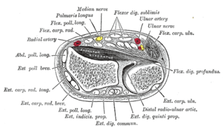
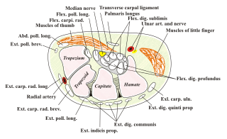
The carpal bones are the eight small bones that make up the wrist (carpus) that connects the hand to the forearm. The terms "carpus" and "carpal" are derived from the Latin carpus and the Greek καρπός (karpós), meaning "wrist". In human anatomy, the main role of the carpal bones is to articulate with the radial and ulnar heads to form a highly mobile condyloid joint, to provide attachments for thenar and hypothenar muscles, and to form part of the rigid carpal tunnel which allows the median nerve and tendons of the anterior forearm muscles to be transmitted to the hand and fingers.

The radial nerve is a nerve in the human body that supplies the posterior portion of the upper limb. It innervates the medial and lateral heads of the triceps brachii muscle of the arm, as well as all 12 muscles in the posterior osteofascial compartment of the forearm and the associated joints and overlying skin.

In human anatomy, the wrist is variously defined as (1) the carpus or carpal bones, the complex of eight bones forming the proximal skeletal segment of the hand; (2) the wrist joint or radiocarpal joint, the joint between the radius and the carpus and; (3) the anatomical region surrounding the carpus including the distal parts of the bones of the forearm and the proximal parts of the metacarpus or five metacarpal bones and the series of joints between these bones, thus referred to as wrist joints. This region also includes the carpal tunnel, the anatomical snuff box, bracelet lines, the flexor retinaculum, and the extensor retinaculum.
The flexor digitorum profundus or flexor digitorum communis profundus is a muscle in the forearm of humans that flexes the fingers. It is considered an extrinsic hand muscle because it acts on the hand while its muscle belly is located in the forearm.
Flexor digitorum superficialis or flexor digitorum communis sublimis is an extrinsic flexor muscle of the fingers at the proximal interphalangeal joints.

The ulnar nerve is a nerve that runs near the ulna, one of the two long bones in the forearm. The ulnar collateral ligament of elbow joint is in relation with the ulnar nerve. The nerve is the largest in the human body unprotected by muscle or bone, so injury is common. This nerve is directly connected to the little finger, and the adjacent half of the ring finger, innervating the palmar aspect of these fingers, including both front and back of the tips, perhaps as far back as the fingernail beds.

The upper limbs or upper extremities are the forelimbs of an upright-postured tetrapod vertebrate, extending from the scapulae and clavicles down to and including the digits, including all the musculatures and ligaments involved with the shoulder, elbow, wrist and knuckle joints. In humans, each upper limb is divided into the shoulder, arm, elbow, forearm, wrist and hand, and is primarily used for climbing, lifting and manipulating objects. In anatomy, just as arm refers to the upper arm, leg refers to the lower leg.
The extensor digitorum muscle is a muscle of the posterior forearm present in humans and other animals. It extends the medial four digits of the hand. Extensor digitorum is innervated by the posterior interosseous nerve, which is a branch of the radial nerve.

In human anatomy, the dorsal interossei (DI) are four muscles in the back of the hand that act to abduct (spread) the index, middle, and ring fingers away from the hand's midline and assist in flexion at the metacarpophalangeal joints and extension at the interphalangeal joints of the index, middle and ring fingers.

In human anatomy, the abductor digiti minimi is a skeletal muscle situated on the ulnar border of the palm of the hand. It forms the ulnar border of the palm and its spindle-like shape defines the hypothenar eminence of the palm together with the skin, connective tissue, and fat surrounding it. Its main function is to pull the little finger away from the other fingers.
In human anatomy, the extensor indicis (proprius) is a narrow, elongated skeletal muscle in the deep layer of the dorsal forearm, placed medial to, and parallel with, the extensor pollicis longus. Its tendon goes to the index finger, which it extends.

The flexor digiti minimi brevis is a hypothenar muscle in the hand that flexes the little finger at the metacarpophalangeal joint. It lies lateral to the abductor digiti minimi when the hand is in anatomical position.
The posterior compartment of the forearm contains twelve muscles which primarily extend the wrist and digits. It is separated from the anterior compartment by the interosseous membrane between the radius and ulna.

The cervical spinal nerve 8 (C8) is a spinal nerve of the cervical segment.
Extensor digitorum brevis manus is an extra or accessory muscle on the backside (dorsum) of the hand. It was first described by Albinus in 1758. The muscles lies in the fourth extensor compartment of the wrist, and is relatively rare. It has a prevalence of 4% in the general population according to a meta-analysis. This muscle is commonly misdiagnosed as a ganglion cyst, synovial nodule or cyst.

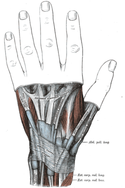
The extrinsic extensor muscles of the hand are located in the back of the forearm and have long tendons connecting them to bones in the hand, where they exert their action. Extrinsic denotes their location outside the hand. Extensor denotes their action which is to extend, or open flat, joints in the hand. They include the extensor carpi radialis longus (ECRL), extensor carpi radialis brevis (ECRB), extensor digitorum (ED), extensor digiti minimi (EDM), extensor carpi ulnaris (ECU), abductor pollicis longus (APL), extensor pollicis brevis (EPB), extensor pollicis longus (EPL), and extensor indicis (EI).

The extensor medii proprius is a rare anatomical variant in the extensor compartment of the forearm. The aberrant muscle is analogous to the extensor indicis with the insertion being the middle finger instead of the index finger.

The extensor indicis et medii communis is a rare anatomical variant in the extensor compartment of forearm. This additional muscle lies in the deep extensor layer next to the extensor indicis proprius and the extensor pollicis longus. The characteristics of this anomalous muscle resemble those of the extensor indicis proprius, with split tendons to the index and the middle finger. This muscle can also be considered as a variation of the aberrant extensor medii proprius.
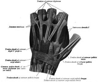
In human anatomy, juncturae tendinum or connexus intertendinei refers to the connective tissues that link the tendons of the extensor digitorum communis, and sometimes, to the tendon of the extensor digiti minimi. Juncturae tendinum are located on the dorsal aspect of the hand in the first, second and third inter-metacarpal spaces proximal to the metacarpophalangeal joint.