Anatomical terms of muscle

Last updated

Anatomical terminology is used to uniquely describe aspects of skeletal muscle, cardiac muscle, and smooth muscle such as their actions, structure, size, and location.

Contents

Types

There are three types of muscle tissue in the body: skeletal, smooth, and cardiac.

Skeletal muscle

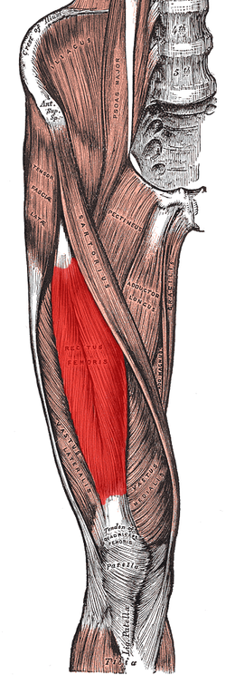
Skeletal muscle, or "voluntary muscle", is a striated muscle tissue that primarily joins to bone with tendons. Skeletal muscle enables movement of bones, and maintains posture. [1] The widest part of a muscle that pulls on the tendons is known as the belly.

Muscle slip

A muscle slip is a slip of muscle that can either be an anatomical variant, [2] or a branching of a muscle as in rib connections of the serratus anterior muscle.

Smooth muscle

Smooth muscle is involuntary and found in parts of the body where it conveys action without conscious intent. The majority of this type of muscle tissue is found in the digestive and urinary systems where it acts by propelling forward food, chyme, and feces in the former and urine in the latter. Other places smooth muscle can be found are within the uterus, where it helps facilitate birth, and the eye, where the pupillary sphincter controls pupil size. [3]

Cardiac muscle

Cardiac muscle is specific to the heart. It is also involuntary in its movement, and is additionally self-excitatory, contracting without outside stimuli. [4]

Actions of skeletal muscle

As well as anatomical terms of motion, which describe the motion made by a muscle, unique terminology is used to describe the action of a set of muscles.

Agonists and antagonists

Agonist muscles and antagonist muscles are muscles that cause or inhibit a movement. [5]

Agonist muscles are also called prime movers since they produce most of the force, and control of an action. [6] Agonists cause a movement to occur through their own activation. [7] For example, the triceps brachii contracts, producing a shortening (concentric) contraction, during the up phase of a push-up (elbow extension). During the down phase of a push-up, the same triceps brachii actively controls elbow flexion while producing a lengthening (eccentric) contraction. It is still the agonist, because while resisting gravity during relaxing, the triceps brachii continues to be the prime mover, or controller, of the joint action.

Another example is the dumb-bell curl at the elbow. The elbow flexor group is the agonist, shortening during the lifting phase (elbow flexion). During the lowering phase the elbow flexor muscles lengthen, remaining the agonists because they are controlling the load and the movement (elbow extension). For both the lifting and lowering phase, the "elbow extensor" muscles are the antagonists (see below). They lengthen during the dumbbell lifting phase and shorten during the dumbbell lowering phase. Here it is important to understand that it is common practice to give a name to a muscle group (e.g. elbow flexors) based on the joint action they produce during a shortening contraction. However, this naming convention does not mean they are only agonists during shortening. This term typically describes the function of skeletal muscles. [8]

Antagonist muscles are simply the muscles that produce an opposing joint torque to the agonist muscles. [9] This torque can aid in controlling a motion. The opposing torque can slow movement down - especially in the case of a ballistic movement. For example, during a very rapid (ballistic) discrete movement of the elbow, such as throwing a dart, the triceps muscles will be activated very briefly and strongly (in a "burst") to rapidly accelerate the extension movement at the elbow, followed almost immediately by a "burst" of activation to the elbow flexor muscles that decelerates the elbow movement to arrive at a quick stop. To use an automotive analogy, this would be similar to pressing the accelerator pedal rapidly and then immediately pressing the brake. Antagonism is not an intrinsic property of a particular muscle or muscle group; it is a role that a muscle plays depending on which muscle is currently the agonist. During slower joint actions that involve gravity, just as with the agonist muscle, the antagonist muscle can shorten and lengthen. Using the example of the triceps brachii during a push-up, the elbow flexor muscles are the antagonists at the elbow during both the up phase and down phase of the movement. During the dumbbell curl, the elbow extensors are the antagonists for both the lifting and lowering phases. [10]

Antagonistic pairs

The antagonistic pair of biceps and triceps working to flex the elbow. Flexion del brazo.png
The antagonistic pair of biceps and triceps working to flex the elbow.

Antagonist and agonist muscles often occur in pairs, called antagonistic pairs. As one muscle contracts, the other relaxes. An example of an antagonistic pair is the biceps and triceps; to contract, the triceps relaxes while the biceps contracts to lift the arm. "Reverse motions" need antagonistic pairs located in opposite sides of a joint or bone, including abductor-adductor pairs and flexor-extensor pairs. These consist of an extensor muscle, which "opens" the joint (by increasing the angle between the two bones) and a flexor muscle, which does the opposite by decreasing the angle between two bones.

However, muscles do not always work this way; sometimes agonists and antagonists contract at the same time to produce force, as per Lombard's paradox. Also, sometimes during a joint action controlled by an agonist muscle, the antagonist will be slightly activated, naturally. This occurs normally and is not considered to be a problem unless it is excessive or uncontrolled and disturbs the control of the joint action. This is called agonist/antagonist co-activation and serves to mechanically stiffen the joint.

Not all muscles are paired in this way. An example of an exception is the deltoid. [11]

Synergists

The biceps flexes the lower arm. The brachioradialis, in the forearm, and brachialis, located deep to the biceps in the upper arm, are both synergists that aid in this motion. Biceps Muscle CNX.jpg
The biceps flexes the lower arm. The brachioradialis, in the forearm, and brachialis, located deep to the biceps in the upper arm, are both synergists that aid in this motion.

Synergist muscles also called fixators, act around a joint to help the action of an agonist muscle. Synergist muscles can also act to counter or neutralize the force of an agonist and are also known as neutralizers when they do this. [12] As neutralizers they help to cancel out or neutralize extra motion produced from the agonists to ensure that the force generated works within the desired plane of motion.

Muscle fibers can only contract up to 40% of their fully stretched length. [ citation needed ] Thus the short fibers of pennate muscles are more suitable where power rather than range of contraction is required. This limitation in the range of contraction affects all muscles, and those that act over several joints may be unable to shorten sufficiently to produce the full range of movement at all of them simultaneously (active insufficiency, e.g., the fingers cannot be fully flexed when the wrist is also flexed). Likewise, the opposing muscles may be unable to stretch sufficiently to allow such movement to take place (passive insufficiency). For both these reasons, it is often essential to use other synergists, in this type of action to fix certain of the joints so that others can be moved effectively, e.g., fixation of the wrist during full flexion of the fingers in clenching the fist. Synergists are muscles that facilitate the fixation action.

There is an important difference between a helping synergist muscle and a true synergist muscle. A true synergist muscle is one that only neutralizes an undesired joint action, whereas a helping synergist is one that neutralizes an undesired action but also assists with the desired action. [ citation needed ]

Neutralizer action

A muscle that fixes or holds a bone so that the agonist can carry out the intended movement is said to have a neutralizing action. A good famous example of this are the hamstrings; the semitendinosus and semimembranosus muscles perform knee flexion and knee internal rotation whereas the biceps femoris carries out knee flexion and knee external rotation. For the knee to flex while not rotating in either direction, all three muscles contract to stabilize the knee while it moves in the desired way.

Composite muscle

Composite or hybrid muscles have more than one set of fibers that perform the same function, and are usually supplied by different nerves for different set of fibers. For example, the tongue itself is a composite muscle made up of various components like longitudinal, transverse, horizontal muscles with different parts innervated having different nerve supply.

Muscle naming

The seven general types of skeletal muscle Fascicle Muscle Shapes.jpg
The seven general types of skeletal muscle

There are a number of terms used in the naming of muscles including those relating to size, shape, action, location, their orientation, and their number of heads.

By size
brevis means short; longus means long; major means large; maximus means largest; minor means small, and minimus smallest. These terms are often used after the particular muscle such as gluteus maximus, and gluteus minimus. [13]
By shape
deltoid means triangular; quadratus means having four sides; rhomboideus means having a rhomboid shape; teres means round or cylindrical, trapezius means having a trapezoid shape, rectus means straight. Examples are the pronator teres, the pronator quadratus and the rectus abdominis. [13]
By action
abductor moving away from the midline; adductor moving towards the midline; depressor moving downwards; elevator moving upwards; flexor moving that decreases an angle; extensor moving that increase an angle or straightens; pronator moving to face down; supinator moving to face upwards; [13] Internal rotator rotating towards the body; external rotator rotating away from the body.

Form

Rectus femoris (in red). Origin includes the anterior inferior iliac spine and part of the acetabulum. Insertion is into the patellar tendon. Action is extension of the knee and flexion of the hip. Rectus femoris.png
Rectus femoris (in red). Origin includes the anterior inferior iliac spine and part of the acetabulum. Insertion is into the patellar tendon. Action is extension of the knee and flexion of the hip.

Insertion and origin

The insertion and origin of a muscle are the two places where it is anchored, one at each end. The connective tissue of the attachment is called an enthesis.

Origin

The origin of a muscle is the bone, typically proximal, which has greater mass and is more stable during a contraction than a muscle's insertion. [14] For example, with the latissimus dorsi muscle, the origin site is the torso, and the insertion is the arm. When this muscle contracts, normally the arm moves due to having less mass than the torso. This is the case when grabbing objects lighter than the body, as in the typical use of a lat pull down machine. This can be reversed however, such as in a chin up where the torso moves up to meet the arm.

The head of a muscle, also called caput musculi is the part at the end of a muscle at its origin, where it attaches to a fixed bone. Some muscles such as the biceps have more than one head.

Insertion

The insertion of a muscle is the structure that it attaches to and tends to be moved by the contraction of the muscle. [15] This may be a bone, a tendon or the subcutaneous dermal connective tissue. Insertions are usually connections of muscle via tendon to bone. [16] The insertion is a bone that tends to be distal, have less mass, and greater motion than the origin during a contraction.

Intrinsic and extrinsic muscles

Intrinsic muscles have their origin in the part of the body that they act on, and are contained within that part. [17] Extrinsic muscles have their origin outside of the part of the body that they act on. [18] Examples are the intrinsic and extrinsic muscles of the tongue, and those of the hand.

Muscle fibers

Different skeletal muscle types:
A: fusiform. B: unipennate. C: bipennate.
(PCS: physiological cross-section) Gray365.png
Different skeletal muscle types:
A: fusiform. B: unipennate. C: bipennate.
(PCS: physiological cross-section)

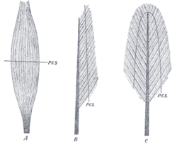
Muscles may also be described by the direction that the muscle fibers run, in their muscle architecture.

State

Hypertrophy and atrophy

Example of an atrophied muscle 1025 Atrophy.png
Example of an atrophied muscle

Hypertrophy is increase in muscle size from an increase in size of individual muscle cells. This usually occurs as a result of exercise.

See also

Related Research Articles

<span class="mw-page-title-main">Arm</span> Proximal part of the free upper limb between the shoulder and the elbow

In human anatomy, the arm refers to the upper limb in common usage, although academically the term specifically means the upper arm between the glenohumeral joint and the elbow joint. The distal part of the upper limb between the elbow and the radiocarpal joint is known as the forearm or "lower" arm, and the extremity beyond the wrist is the hand.

<span class="mw-page-title-main">Ulna</span> Medial bone from forearm

The ulna or ulnal bone is a long bone found in the forearm that stretches from the elbow to the wrist, and when in anatomical position, is found on the medial side of the forearm. That is, the ulna is on the same side of the forearm as the little finger. It runs parallel to the radius, the other long bone in the forearm. The ulna is longer and the radius is shorter, but the radius is thicker and the ulna is thinner. Therefore, the ulna is considered to be the smaller bone of the two bones in the lower arm. The corresponding bone in the lower leg is the fibula.

<span class="mw-page-title-main">Humerus</span> Long bone of the upper arm

The humerus is a long bone in the arm that runs from the shoulder to the elbow. It connects the scapula and the two bones of the lower arm, the radius and ulna, and consists of three sections. The humeral upper extremity consists of a rounded head, a narrow neck, and two short processes. The body is cylindrical in its upper portion, and more prismatic below. The lower extremity consists of 2 epicondyles, 2 processes, and 3 fossae. As well as its true anatomical neck, the constriction below the greater and lesser tubercles of the humerus is referred to as its surgical neck due to its tendency to fracture, thus often becoming the focus of surgeons.

<span class="mw-page-title-main">Biceps</span> Muscle on the front of the upper arm

The biceps or biceps brachii are a large muscle that lies on the front of the upper arm between the shoulder and the elbow. Both heads of the muscle arise on the scapula and join to form a single muscle belly which is attached to the upper forearm. While the biceps crosses both the shoulder and elbow joints, its main function is at the elbow where it flexes the forearm and supinates the forearm. Both these movements are used when opening a bottle with a corkscrew: first biceps screws in the cork (supination), then it pulls the cork out (flexion).

<span class="mw-page-title-main">Brachioradialis</span> Muscle of the upper limb

The brachioradialis is a muscle of the forearm that flexes the forearm at the elbow. It is also capable of both pronation and supination, depending on the position of the forearm. It is attached to the distal styloid process of the radius by way of the brachioradialis tendon, and to the lateral supracondylar ridge of the humerus.

<span class="mw-page-title-main">Brachialis muscle</span> Flexor muscle in the upper arm

The brachialis is a muscle in the upper arm that flexes the elbow. It lies beneath the biceps brachii, and makes up part of the floor of the region known as the cubital fossa. It originates from the anterior aspect of the distal humerus; it inserts onto the tuberosity of the ulna. It is innervated by the musculocutaneous nerve, and commonly also receives additional innervation from the radial nerve. The brachialis is the prime mover of elbow flexion generating about 50% more power than the biceps.

<span class="mw-page-title-main">Radius (bone)</span> One of the two long bones of the forearm

The radius or radial bone is one of the two large bones of the forearm, the other being the ulna. It extends from the lateral side of the elbow to the thumb side of the wrist and runs parallel to the ulna. The ulna is longer than the radius, but the radius is thicker. The radius is a long bone, prism-shaped and slightly curved longitudinally.

<span class="mw-page-title-main">Upper limb</span> Consists of the arm, forearm, and hand

The upper limbs or upper extremities are the forelimbs of an upright-postured tetrapod vertebrate, extending from the scapulae and clavicles down to and including the digits, including all the musculatures and ligaments involved with the shoulder, elbow, wrist and knuckle joints. In humans, each upper limb is divided into the arm, forearm and hand, and is primarily used for climbing, lifting and manipulating objects.

<span class="mw-page-title-main">Triceps</span> Muscle on the back of the upper arm

The triceps, or triceps brachii, is a large muscle on the back of the upper limb of many vertebrates. It consists of 3 parts: the medial, lateral, and long head. It is the muscle principally responsible for extension of the elbow joint.

René Descartes (1596–1650) was one of the first to conceive a model of reciprocal innervation as the principle that provides for the control of agonist and antagonist muscles. Reciprocal innervation describes skeletal muscles as existing in antagonistic pairs, with contraction of one muscle producing forces opposite to those generated by contraction of the other. For example, in the human arm, the triceps acts to extend the lower arm outward while the biceps acts to flex the lower arm inward. To reach optimum efficiency, contraction of opposing muscles must be inhibited while muscles with the desired action are excited. This reciprocal innervation occurs so that the contraction of a muscle results in the simultaneous relaxation of its corresponding antagonist.

Reciprocal inhibition describes the relaxation of muscles on one side of a joint to accommodate contraction on the other side. In some allied health disciplines, this is known as reflexive antagonism. The central nervous system sends a message to the agonist muscle to contract. The tension in the antagonist muscle is activated by impulses from motor neurons, causing it to relax.

The pronator teres is a muscle that, along with the pronator quadratus, serves to pronate the forearm. It has two origins, at the medial humeral supracondylar ridge and the ulnar tuberosity, and inserts near the middle of the radius.

<span class="mw-page-title-main">Stretch reflex</span> Muscle contraction in response to stretching

The stretch reflex, or more accurately "muscle stretch reflex", is a muscle contraction in response to stretching a muscle. The function of the reflex is generally thought to be maintaining the muscle at a constant length but the response is often coordinated across multiple muscles and even joints. The older term deep tendon reflex is now criticized as misleading. Tendons have little to do with the response, and some muscles with stretch reflexes have no tendons. Rather, muscle spindles detect a stretch and convey the information to the central nervous system.

<span class="mw-page-title-main">Fascial compartments of arm</span> Anatomical compartments

The fascial compartments of arm refers to the specific anatomical term of the compartments within the upper segment of the upper limb of the body. The upper limb is divided into two segments, the arm and the forearm. Each of these segments is further divided into two compartments which are formed by deep fascia – tough connective tissue septa (walls). Each compartment encloses specific muscles and nerves.

<span class="mw-page-title-main">Elbow</span> Joint between the upper and lower parts of the arm

The elbow is the region between the upper arm and the forearm that surrounds the elbow joint. The elbow includes prominent landmarks such as the olecranon, the cubital fossa, and the lateral and the medial epicondyles of the humerus. The elbow joint is a hinge joint between the arm and the forearm; more specifically between the humerus in the upper arm and the radius and ulna in the forearm which allows the forearm and hand to be moved towards and away from the body. The term elbow is specifically used for humans and other primates, and in other vertebrates forelimb plus joint is used.

Upper-limb surgery in tetraplegia includes a number of surgical interventions that can help improve the quality of life of a patient with tetraplegia.

<span class="mw-page-title-main">Anatomical terminology</span> Scientific terminology used by anatomists, zoologists, and health professionals

Anatomical terminology is a form of scientific terminology used by anatomists, zoologists, and health professionals such as doctors, physicians, and pharmacists.

<span class="mw-page-title-main">Stay apparatus</span>

The stay apparatus is an arrangement of muscles, tendons and ligaments that work together so that an animal can remain standing with virtually no muscular effort. It is best known as the mechanism by which horses can enter a light sleep while still standing up. The effect is that an animal can distribute its weight on three limbs while resting a fourth in a flexed, non-weight bearing position. The animal can periodically shift its weight to rest a different leg and thus all limbs are able to be individually rested, reducing overall wear and tear. The relatively slim legs of certain large mammals such as horses and cows would be subject to dangerous levels of fatigue if not for the stay apparatus.

References

PD-icon.svgThis article incorporates text in the public domain from the 20th edition of Gray's Anatomy (1918)

  1. Skeletal Muscle
  2. Stimec, Bojan V.; Dash, Jérémy; Assal, Mathieu; Stern, Richard; Fasel, Jean H. D. (1 May 2018). "Additional muscular slip of the flexor digitorum longus muscle to the fifth toe". Surgical and Radiologic Anatomy. 40 (5): 533–535. doi:10.1007/s00276-018-1991-7. PMID   29473094. S2CID   3456242 . Retrieved 13 May 2021.
  3. Smooth Muscle
  4. Cardiac Muscle
  5. "Interactions of skeletal muscles their fascicle arrangement and their lever-systems". Archived from the original on 23 March 2022. Retrieved 10 May 2021.
  6. Saladin, Kenneth S. (2011). Human anatomy (3rd ed.). New York: McGraw-Hill. pp. 236–241. ISBN   9780071222075.
  7. Taber 2001, pp. "Agonist".
  8. Baechle, Thomas (2008). Essentials of Strength Training and Conditioning. USA: National Strength and Conditioning Association. ISBN   978-0-7360-8465-9.
  9. Taber 2001, pp. "Antagonist".
  10. Walker, H. Kenneth (1990), Walker, H. Kenneth; Hall, W. Dallas; Hurst, J. Willis (eds.), "Deep Tendon Reflexes", Clinical Methods: The History, Physical, and Laboratory Examinations (3rd ed.), Boston: Butterworths, ISBN   978-0-409-90077-4, PMID   21250237 , retrieved 2024-02-19
  11. Purves, D; Augustine, GJ (2001). "Neural Circuits". NCBI. Sinauer Association.
  12. "9.6C: How Skeletal Muscles Produce Movements". Medicine LibreTexts. 19 July 2018. Retrieved 8 May 2021.
  13. 1 2 3 Saladin, Kenneth S. (2011). Human anatomy (3rd ed.). New York: McGraw-Hill. p. 265. ISBN   9780071222075.
  14. OED 1989, "origin".
  15. Taber 2001, "insertion".
  16. Martini, Frederic; William C. Ober; Claire W. Garrison; Kathleen Welch; Ralph T. Hutchings (2001). Fundamentals of Anatomy and Physiology, 5th Ed. Prentice Hall. ISBN   0130172928.
  17. "Definition of INTRINSIC". www.merriam-webster.com. Retrieved 7 May 2021.
  18. "Definition of EXTRINSIC". www.merriam-webster.com. Retrieved 7 May 2021.
  19. Taber 2001, "Fusiform".
Books