Muscles of the hand

Last updated
Muscles of the hand
Wrist and hand deeper palmar dissection.svg
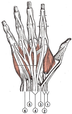
Muscles and other structures of wrist and palm
Details
Origin Upper extremity
Insertion Hand and fingers
Nerve radial, median, and ulnar nerves (from C5T1)
Actions Flexion, extension, adduction, abduction of the hand and fingers and oppossion of the thumb
Identifiers
FMA 42368
Anatomical terms of muscle

The muscles of the hand are the skeletal muscles responsible for the movement of the hand and fingers. The muscles of the hand can be subdivided into two groups: the extrinsic and intrinsic muscle groups. The extrinsic muscle groups are the long flexors and extensors. They are called extrinsic because the muscle belly is located on the forearm. The intrinsic group are the smaller muscles located within the hand itself. The muscles of the hand are innervated by the radial, median, and ulnar nerves from the brachial plexus. [1]

Contents

Intrinsic

The intrinsic muscle groups are the thenar (thumb) and hypothenar (little finger) muscles; the interossei muscles (four dorsally and three volarly) originating between the metacarpal bones; and the lumbrical muscles arising from the deep flexor (and which are special because they have no bony origin) to insert on the dorsal extensor hood mechanism. Palmaris brevis which is a superficial muscle and adductor pollicis are also intrinsic muscles. [2]

Extrinsic

Extensor compartments of wrist (back of hand) Wrist extensor compartments (numbered).PNG
Extensor compartments of wrist (back of hand)

The fingers have two long flexors, located on the underside of the forearm. They insert by tendons to the phalanges of the fingers. The deep flexor attaches to the distal phalanx, and the superficial flexor attaches to the middle phalanx. The flexors allow for the actual bending of the fingers. The thumb has one long flexor and a short flexor in the thenar muscle group. The human thumb also has other muscles in the thenar group (opponens and abductor brevis muscle), moving the thumb in opposition, making grasping possible.

The extensors are located on the back of the forearm and are connected in a more complex way than the flexors to the dorsum of the fingers. The tendons unite with the interosseous and lumbrical muscles to form the extensorhood mechanism. The primary function of the extensors is to straighten out the digits. The thumb has two extensors in the forearm; the tendons of these form the anatomical snuff box. Also, the index finger and the little finger have an extra extensor, used, for instance, for pointing. The extensors are situated within 6 separate compartments.

Compartment 1 (Most radial)Compartment 2Compartment 3Compartment 4Compartment 5Compartment 6 (Most ulnar)
Abductor pollicis longus Extensor carpi radialis longus Extensor pollicis longus Extensor indicis Extensor digiti minimi Extensor carpi ulnaris
Extensor pollicis brevis Extensor carpi radialis brevis Extensor digitorum communis

The first four compartments are located in the grooves present on the dorsum of inferior side of radius, while the 5th compartment is in between radius and ulna. The 6th compartment is in the groove on the dorsum of inferior side of ulna.

Nerve supply

The muscles of the hand are innervated by the radial, median, and ulnar nerves.

The radial nerve innervates the finger extensors and the thumb abductor; that is, the muscles that extend at the wrist and metacarpophalangeal joints (knuckles) and abduct and extend the thumb. The median nerve innervates the flexors of the wrist and digits, the abductors and opponens of the thumb, the first and second lumbricals. The ulnar nerve innervates the remaining intrinsic muscles of the hand. [3] [4]

All muscles of the hand are innervated by the brachial plexus (C5T1) and can be classified by innervation: [1] [4] [5]

NerveMuscles
RadialExtensors: carpi radialis longus and brevis, digitorum, digiti minimi, carpi ulnaris, pollicis longus and brevis, and indicis.
Other: abductor pollicis longus.
MedianFlexors: carpi radialis, pollicis longus, digitorum profundus (half), superficialis, and pollicis brevis (superficial head).
Other: palmaris longus. abductor pollicis brevis, opponens pollicis, and first and second lumbricals.
Ulnar Flexor carpi ulnaris, flexor digitorum profundus (half), palmaris brevis, flexor digiti minimi, abductor digiti minimi, opponens digiti minimi, adductor pollicis, flexor pollicis brevis (deep head), palmar and dorsal interossei, and third and fourth lumbricals.

See also

Related Research Articles

<span class="mw-page-title-main">Finger</span> Organ of manipulation and sensation found in the hands of humans and other primates

A finger is a prominent digit on the forelimbs of most tetrapod vertebrate animals, especially those with prehensile extremities such as humans and other primates. Most tetrapods have five digits (pentadactyly), and short digits are typically referred to as toes, while those that are notably elongated are called fingers. In humans, the fingers are flexibly articulated and opposable, serving as an important organ of tactile sensation and fine movements, which are crucial to the dexterity of the hands and the ability to grasp and manipulate objects.

<span class="mw-page-title-main">Thumb</span> First digit of the hand

The thumb is the first digit of the hand, next to the index finger. When a person is standing in the medical anatomical position, the thumb is the outermost digit. The Medical Latin English noun for thumb is pollex, and the corresponding adjective for thumb is pollical.

<span class="mw-page-title-main">Radial nerve</span> Nerve in the human body that supplies the posterior portion of the upper limb

The radial nerve is a nerve in the human body that supplies the posterior portion of the upper limb. It innervates the medial and lateral heads of the triceps brachii muscle of the arm, as well as all 12 muscles in the posterior osteofascial compartment of the forearm and the associated joints and overlying skin.

<span class="mw-page-title-main">Median nerve</span> Nerve of the upper limb

The median nerve is a nerve in humans and other animals in the upper limb. It is one of the five main nerves originating from the brachial plexus.

The flexor digitorum profundus is a muscle in the forearm of humans that flexes the fingers. It is considered an extrinsic hand muscle because it acts on the hand while its muscle belly is located in the forearm.

<span class="mw-page-title-main">Thenar eminence</span> Mound at the base of the thumb formed by the intrinsic group of muscles

The thenar eminence is the mound formed at the base of the thumb on the palm of the hand by the intrinsic group of muscles of the thumb. The skin overlying this region is the area stimulated when trying to elicit a palmomental reflex. The word thenar comes from Ancient Greek θέναρ (thenar) 'palm of the hand'.

The forearm is the region of the upper limb between the elbow and the wrist. The term forearm is used in anatomy to distinguish it from the arm, a word which is used to describe the entire appendage of the upper limb, but which in anatomy, technically, means only the region of the upper arm, whereas the lower "arm" is called the forearm. It is homologous to the region of the leg that lies between the knee and the ankle joints, the crus.

<span class="mw-page-title-main">Ulnar nerve</span> Nerve which runs near the ulna bone

In human anatomy, the ulnar nerve is a nerve that runs near the ulna bone. The ulnar collateral ligament of elbow joint is in relation with the ulnar nerve. The nerve is the largest in the human body unprotected by muscle or bone, so injury is common. This nerve is directly connected to the little finger, and the adjacent half of the ring finger, innervating the palmar aspect of these fingers, including both front and back of the tips, perhaps as far back as the fingernail beds.

<span class="mw-page-title-main">Wrist drop</span> Medical condition

Wrist drop is a medical condition in which the wrist and the fingers cannot extend at the metacarpophalangeal joints. The wrist remains partially flexed due to an opposing action of flexor muscles of the forearm. As a result, the extensor muscles in the posterior compartment remain paralyzed.

<span class="mw-page-title-main">Upper limb</span> Consists of the arm, forearm, and hand

The upper limbs or upper extremities are the forelimbs of an upright-postured tetrapod vertebrate, extending from the scapulae and clavicles down to and including the digits, including all the musculatures and ligaments involved with the shoulder, elbow, wrist and knuckle joints. In humans, each upper limb is divided into the arm, forearm and hand, and is primarily used for climbing, lifting and manipulating objects.

<span class="mw-page-title-main">Extensor digitorum muscle</span> Muscle of the posterior forearm

The extensor digitorum muscle is a muscle of the posterior forearm present in humans and other animals. It extends the medial four digits of the hand. Extensor digitorum is innervated by the posterior interosseous nerve, which is a branch of the radial nerve.

In human anatomy, the abductor pollicis longus (APL) is one of the extrinsic muscles of the hand. Its major function is to abduct the thumb at the wrist. Its tendon forms the anterior border of the anatomical snuffbox.

<span class="mw-page-title-main">Adductor pollicis muscle</span> Muscle in the thenar compartment

In human anatomy, the adductor pollicis muscle is a muscle in the hand that functions to adduct the thumb. It has two heads: transverse and oblique.

<span class="mw-page-title-main">Opponens pollicis muscle</span> Muscle in the thenar compartment

The opponens pollicis is a small, triangular muscle in the hand, which functions to oppose the thumb. It is one of the three thenar muscles. It lies deep to the abductor pollicis brevis and lateral to the flexor pollicis brevis.

The posterior compartment of the forearm contains twelve muscles which primarily extend the wrist and digits. It is separated from the anterior compartment by the interosseous membrane between the radius and ulna.

<span class="mw-page-title-main">Ulnar claw</span> Deformity of the hand that develops due to ulnar nerve damage

An ulnar claw, also known as claw hand or ‘Spinster’s Claw’, is a deformity or an abnormal attitude of the hand that develops due to ulnar nerve damage causing paralysis of the lumbricals. A claw hand presents with a hyperextension at the metacarpophalangeal joints and flexion at the proximal and distal interphalangeal joints of the 4th and 5th fingers. The patients with this condition can make a full fist but when they extend their fingers, the hand posture is referred to as claw hand. The ring- and little finger can usually not fully extend at the proximal interphalangeal joint (PIP).

<span class="mw-page-title-main">Hand</span> Extremity at the end of an arm or forelimb

A hand is a prehensile, multi-fingered appendage located at the end of the forearm or forelimb of primates such as humans, chimpanzees, monkeys, and lemurs. A few other vertebrates such as the koala are often described as having "hands" instead of paws on their front limbs. The raccoon is usually described as having "hands" though opposable thumbs are lacking.

<span class="mw-page-title-main">Median nerve palsy</span> Medical condition

Injuries to the arm, forearm or wrist area can lead to various nerve disorders. One such disorder is median nerve palsy. The median nerve controls the majority of the muscles in the forearm. It controls abduction of the thumb, flexion of hand at wrist, flexion of digital phalanx of the fingers, is the sensory nerve for the first three fingers, etc. Because of this major role of the median nerve, it is also called the eye of the hand. If the median nerve is damaged, the ability to abduct and oppose the thumb may be lost due to paralysis of the thenar muscles. Various other symptoms can occur which may be repaired through surgery and tendon transfers. Tendon transfers have been very successful in restoring motor function and improving functional outcomes in patients with median nerve palsy.

<span class="mw-page-title-main">Extrinsic extensor muscles of the hand</span>

The extrinsic extensor muscles of the hand are located in the back of the forearm and have long tendons connecting them to bones in the hand, where they exert their action. Extrinsic denotes their location outside the hand. Extensor denotes their action which is to extend, or open flat, joints in the hand. They include the extensor carpi radialis longus (ECRL), extensor carpi radialis brevis (ECRB), extensor digitorum (ED), extensor digiti minimi (EDM), extensor carpi ulnaris (ECU), abductor pollicis longus (APL), extensor pollicis brevis (EPB), extensor pollicis longus (EPL), and extensor indicis (EI).

<span class="mw-page-title-main">Muscles of the thumb</span>

The muscles of the thumb are nine skeletal muscles located in the hand and forearm. The muscles allow for flexion, extension, adduction, abduction and opposition of the thumb. The muscles acting on the thumb can be divided into two groups: The extrinsic hand muscles, with their muscle bellies located in the forearm, and the intrinsic hand muscles, with their muscles bellies located in the hand proper.

References

  1. 1 2 Ross & Lamperti 2006 , p. 257
  2. "Medical mnemonics". LifeHugger. Archived from the original on 2011-07-13. Retrieved 2009-12-19.
  3. Jones & Lederman 2006 , pp. 16–18
  4. 1 2 Palazzo, J. J.; Galloway, K. (2017-01-01), Placzek, Jeffrey D.; Boyce, David A. (eds.), "Chapter 53 - Nerve Entrapments of the Wrist and Hand", Orthopaedic Physical Therapy Secrets (Third Edition), Elsevier, pp. 429–436, doi:10.1016/b978-0-323-28683-1.00053-9, ISBN   978-0-323-28683-1 , retrieved 2020-10-24
  5. Jacobson, Mark D.; Raab, Rajnik; Fazeli, Babak M.; Abrams, Reid A.; Botte, Michael J.; Lieber, Richard L. (September 1992). "Architectural design of the human intrinsic hand muscles" . The Journal of Hand Surgery. 17 (5): 804–809. doi:10.1016/0363-5023(92)90446-v. ISSN   0363-5023. PMID   1401784.