Metacarpophalangeal joint

Last updated
Metacarpophalangeal joint
Grant 1962 104 MCP.png
The palmar aspect of the hand showing the epiphyses of the hand exploded. MCP joints in red.
DIP, PIP and MCP joints of hand.jpg
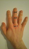
The DIP, PIP and MCP joints of the hand: MetaCarpoPhalangeal joints, and the interphalangeal joints of the hand:
  • Distal InterPhalangeal
  • Proximal InterPhalangeal
Details
System 099
Identifiers
Latin articulationes metacarpophalangeae
MeSH D008662
TA98 A03.5.11.501
TA2 1835
FMA 35246
Anatomical terminology

The metacarpophalangeal joints (MCP) are situated between the metacarpal bones and the proximal phalanges of the fingers. [1] These joints are of the condyloid kind, formed by the reception of the rounded heads of the metacarpal bones into shallow cavities on the proximal ends of the proximal phalanges. [1] Being condyloid, they allow the movements of flexion, extension, abduction, adduction and circumduction at the joint. [1]

Contents

Structure

Ligaments

Gray337.png
Metacarpophalangeal articulation and articulations of digit. Palmar aspect.
Gray338.png
Metacarpophalangeal articulation and articulations of digit. Ulnar aspect.

Each joint has:

Dorsal surfaces

The dorsal surfaces of these joints are covered by the expansions of the Extensor tendons, together with some loose areolar tissue which connects the deep surfaces of the tendons to the bones.

Function

The movements which occur in these joints are flexion, extension, adduction, abduction, and circumduction; the movements of abduction and adduction are very limited, and cannot be performed while the fingers form a fist. [2]

The muscles of flexion and extension are as follows:

LocationFlexionExtension
fingers Flexor digitorum superficialis and profundus, lumbricals, and interossei, assisted in the case of the little finger by the flexor digiti minimi brevis extensor digitorum communis, extensor indicis proprius, and extensor digiti minimi muscle
thumb flexor pollicis longus and brevis extensor pollicis longus and brevis

Clinical significance

Arthritis of the MCP is a distinguishing feature of rheumatoid arthritis, as opposed to the distal interphalangeal joint in osteoarthritis.

Other animals

In many quadrupeds, particularly horses and other larger animals, the metacarpophalangeal joint is referred to as the "fetlock". This term is translated literally as "foot-lock". In fact, although the term fetlock does not specifically apply to other species' metacarpophalangeal joints (for instance, humans), the "second" or "mid-finger" knuckle of the human hand does anatomically correspond to the fetlock on larger quadrupeds. For lack of a better term, the shortened name may seem more practical.

Related Research Articles

Carpal bones Eight small bones that make up the wrist (or carpus) that connects the hand to the forearm

The carpal bones are the eight small bones that make up the wrist that connects the hand to the forearm. The term "carpus" is derived from the Latin carpus and the Greek καρπός (karpós), meaning "wrist". In human anatomy, the main role of the wrist is to facilitate effective positioning of the hand and powerful use of the extensors and flexors of the forearm, and the mobility of individual carpal bones increase the freedom of movements at the wrist.

Wrist Part of the arm between the lower arm and the hand

In human anatomy, the wrist is variously defined as (1) the carpus or carpal bones, the complex of eight bones forming the proximal skeletal segment of the hand; (2) the wrist joint or radiocarpal joint, the joint between the radius and the carpus and; (3) the anatomical region surrounding the carpus including the distal parts of the bones of the forearm and the proximal parts of the metacarpus or five metacarpal bones and the series of joints between these bones, thus referred to as wrist joints. This region also includes the carpal tunnel, the anatomical snuff box, bracelet lines, the flexor retinaculum, and the extensor retinaculum.

Metacarpal bones

In human anatomy, the metacarpal bones or metacarpus form the intermediate part of the skeletal hand located between the phalanges of the fingers and the carpal bones of the wrist, which forms the connection to the forearm. The metacarpal bones are analogous to the metatarsal bones in the foot.

Upper limb

The upper limbs or upper extremities are the forelimbs of an upright-postured tetrapod vertebrate, extending from the scapulae and clavicles down to and including the digits, including all the musculatures and ligaments involved with the shoulder, elbow, wrist and knuckle joints. In humans, each upper limb is divided into the arm, forearm and hand, and is primarily used for climbing, lifting and manipulating objects.

Phalanx bone Digital bone in the hands and feet of most vertebrates

The phalanges are digital bones in the hands and feet of most vertebrates. In primates, the thumbs and big toes have two phalanges while the other digits have three phalanges. The phalanges are classed as long bones.

Extensor digitorum muscle

The extensor digitorum muscle is a muscle of the posterior forearm present in humans and other animals. It extends the medial four digits of the hand. Extensor digitorum is innervated by the posterior interosseous nerve, which is a branch of the radial nerve.

Fetlock

Fetlock is the common name in horses, large animals, and sometimes dogs for the metacarpophalangeal and metatarsophalangeal joints.

Carpometacarpal joint

The carpometacarpal (CMC) joints are five joints in the wrist that articulate the distal row of carpal bones and the proximal bases of the five metacarpal bones.

Dorsal interossei of the hand

In human anatomy, the dorsal interossei (DI) are four muscles in the back of the hand that act to abduct (spread) the index, middle, and ring fingers away from hand's midline and assist in flexion at the metacarpophalangeal joints and extension at the interphalangeal joints of the index, middle and ring fingers.

Abductor digiti minimi muscle of hand

In human anatomy, the abductor digiti minimi is a skeletal muscle situated on the ulnar border of the palm of the hand. It forms the ulnar border of the palm and its spindle-like shape defines the hypothenar eminence of the palm together with the skin, connective tissue, and fat surrounding it. Its main function is to pull the little finger away from the other fingers.

Interphalangeal joints of the hand

The interphalangeal joints of the hand also known as Bitals, are the hinge joints between the phalanges of the fingers that provide flexion towards the palm of the hand.

Metatarsophalangeal joints

The metatarsophalangeal joints, also informally known as toe knuckles, are the joints between the metatarsal bones of the foot and the proximal bones of the toes. They are condyloid joints, meaning that an elliptical or rounded surface comes close to a shallow cavity.

Condyloid joint

A condyloid joint is an ovoid articular surface, or condyle that is received into an elliptical cavity. This permits movement in two planes, allowing flexion, extension, adduction, abduction, and circumduction.

Deep transverse metacarpal ligament Ligament that connects the palmar surfaces of the heads of the second to fifth metacarpal bones

The deep transverse metacarpal ligament is a narrow fibrous band which runs across the palmar surfaces of the heads of the second, third, fourth and fifth metacarpal bones, connecting them together.

Interphalangeal joints of the foot

The interphalangeal joints of the foot are between the phalanx bones of the toes in the feet.

Midcarpal joint

The midcarpal joint is formed by the scaphoid, lunate, and triquetral bones in the proximal row, and the trapezium, trapezoid, capitate, and hamate bones in the distal row. The distal pole of the scaphoid articulates with two trapezial bones as a gliding type of joint. The proximal end of the scaphoid combines with the lunate and triquetrum to form a deep concavity that articulates with the convexity of the combined capitate and hamate in a form of diarthrodial, almost condyloid joint.

Collateral ligaments of metacarpophalangeal joints

In human anatomy, the radial (RCL) and ulnar (UCL) collateral ligaments of the metacarpophalangeal joints (MCP) of the hand are the primary stabilisers of the MCP joints. They have two parts: the cord-like collateral ligaments proper located more dorsally and the accessory collateral ligaments located more volarly. They enable us to spread our fingers with an open hand but not with the hand closed into a fist.

Palmar plate

In the human hand, palmar or volar plates are found in the metacarpophalangeal (MCP) and interphalangeal (IP) joints, where they reinforce the joint capsules, enhance joint stability, and limit hyperextension. The plates of the MCP and IP joints are structurally and functionally similar, except that in the MCP joints they are interconnected by a deep transverse ligament. In the MCP joints, they also indirectly provide stability to the longitudinal palmar arches of the hand. The volar plate of the thumb MCP joint has a transverse longitudinal rectangular shape, shorter than those in the fingers.

Hand Extremity at the end of an arm or forelimb

A hand is a prehensile, multi-fingered appendage located at the end of the forearm or forelimb of primates such as humans, chimpanzees, monkeys, and lemurs. A few other vertebrates such as the koala are often described as having "hands" instead of paws on their front limbs. The raccoon is usually described as having "hands" though opposable thumbs are lacking.

Extrinsic extensor muscles of the hand

The extrinsic extensor muscles of the hand are located in the back of the forearm and have long tendons connecting them to bones in the hand, where they exert their action. Extrinsic denotes their location outside the hand. Extensor denotes their action which is to extend, or open flat, joints in the hand. They include the extensor carpi radialis longus (ECRL), extensor carpi radialis brevis (ECRB), extensor digitorum (ED), extensor digiti minimi (EDM), extensor carpi ulnaris (ECU), abductor pollicis longus (APL), extensor pollicis brevis (EPB), extensor pollicis longus (EPL), and extensor indicis (EI).

References

PD-icon.svgThis article incorporates text in the public domain from page 332 of the 20th edition of Gray's Anatomy (1918)

  1. 1 2 3 Drake, Vogl and Mitchell (2015). Grey's Anatomy for Students, 3rd Edition. Churchill Livingstone, Elsevier. p. 796. ISBN   9780702051319.
  2. Gray's Anatomy (1918), see infobox