Inferior transverse ligament of scapula | |
---|---|
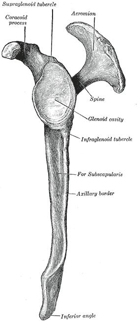
![]() Left scapula. Lateral view. (Ligament not visible, but sites of attachments of the ligament can be seen.) | |
Details | |
From | Spine of scapula |
To | Glenoid cavity |
Identifiers | |
Latin | ligamentum transversum scapulae inferius |
TA98 | A03.5.01.004 |
TA2 | 1742 |
FMA | 25978 |
Anatomical terminology |
The inferior transverse ligament (spinoglenoid ligament [1] ) is a weak membranous band, situated behind the neck of the scapula and stretching from the lateral border of the spine to the margin of the glenoid cavity.
It forms an arch under which the transverse scapular vessels and suprascapular nerve enter the infraspinatous fossa.
In anatomy, the atlas (C1) is the most superior (first) cervical vertebra of the spine and is located in the neck.
The clavicle, collarbone, or keybone is a slender, S-shaped long bone approximately 6 inches (15 cm) long that serves as a strut between the shoulder blade and the sternum (breastbone). There are two clavicles, one on each side of the body. The clavicle is the only long bone in the body that lies horizontally. Together with the shoulder blade, it makes up the shoulder girdle. It is a palpable bone and, in people who have less fat in this region, the location of the bone is clearly visible. It receives its name from Latin clavicula 'little key' because the bone rotates along its axis like a key when the shoulder is abducted. The clavicle is the most commonly fractured bone. It can easily be fractured by impacts to the shoulder from the force of falling on outstretched arms or by a direct hit.
The scapula, also known as the shoulder blade, is the bone that connects the humerus with the clavicle. Like their connected bones, the scapulae are paired, with each scapula on either side of the body being roughly a mirror image of the other. The name derives from the Classical Latin word for trowel or small shovel, which it was thought to resemble.
The coracoid process is a small hook-like structure on the lateral edge of the superior anterior portion of the scapula. Pointing laterally forward, it, together with the acromion, serves to stabilize the shoulder joint. It is palpable in the deltopectoral groove between the deltoid and pectoralis major muscles.
The human shoulder is made up of three bones: the clavicle (collarbone), the scapula, and the humerus as well as associated muscles, ligaments and tendons.
The levator scapulae is a slender skeletal muscle situated at the back and side of the neck. It originates from the transverse processes of the four uppermost cervical vertebrae; it inserts onto the upper portion of the medial border of the scapula. It is innervated by the cervical nerves C3-C4, and frequently also by the dorsal scapular nerve. As the Latin name suggests, its main function is to lift the scapula.
The rhomboid major is a skeletal muscle of the back that connects the scapula with the vertebrae of the spinal column. It originates from the spinous processes of the thoracic vertebrae T2–T5 and supraspinous ligament; it inserts onto the lower portion of the medial border of the scapula. It acts together with the rhomboid minor to keep the scapula pressed against thoracic wall and to retract the scapula toward the vertebral column.
In human anatomy, the rhomboid minor is a small skeletal muscle of the back that connects the scapula to the vertebrae of the spinal column. It arises from the nuchal ligament, and the 7th cervical and 1st thoracic vertebrae and intervening supraspinous ligaments; it inserts onto the medial border of the scapula. It is innervated by the dorsal scapular nerve.
The suprascapular nerve is a mixed nerve that branches from the upper trunk of the brachial plexus. It is derived from the ventral rami of cervical nerves C5-C6. It provides motor innervation to the supraspinatus muscle, and the infraspinatus muscle.
The serratus anterior is a muscle of the chest. It originates at the side of the chest from the upper 8 or 9 ribs; it inserts along the entire length of the anterior aspect of the medial border of the scapula. It is innervated by the long thoracic nerve from the brachial plexus. The serratus anterior acts to pull the scapula forward around the thorax.
The shoulder joint is structurally classified as a synovial ball-and-socket joint and functionally as a diarthrosis and multiaxial joint. It involves an articulation between the glenoid fossa of the scapula and the head of the humerus. Due to the very loose joint capsule ,that gives a limited interface of the humerus and scapula, it is the most mobile joint of the human body.
The suprascapular artery is a branch of the thyrocervical trunk on the neck.
The coracoclavicular ligament is a ligament of the shoulder. It connects the clavicle to the coracoid process of the scapula.
The sternoclavicular joint or sternoclavicular articulation is a synovial saddle joint between the manubrium of the sternum, and the clavicle, and the first costal cartilage. The joint possesses a joint capsule, and an articular disc, and is reinforced by multiple ligaments.
The supraspinous fossa of the posterior aspect of the scapula is smaller than the infraspinous fossa, concave, smooth, and broader at its vertebral than at its humeral end. Its medial two-thirds give origin to the Supraspinatus.
The superior transverse ligament converts the suprascapular notch into a foramen or opening.
A transverse ligament is a ligament on a transverse plane, orthogonal to the anteroposterior or oral-aboral axiscan of the body.
The suprascapular notch is a notch in the superior border of the scapula, just medial to the base of the coracoid process. It is converted into the suprascapular canal by the suprascapular ligament.
The porta hepatis or transverse fissure of the liver is a short but deep fissure, about 5 cm long, extending transversely beneath the left portion of the right lobe of the liver, nearer its posterior surface than its anterior border.
This article incorporates text in the public domain from page 317 of the 20th edition of Gray's Anatomy (1918)