
The carpal bones are the eight small bones that make up the wrist (carpus) that connects the hand to the forearm. The term "carpus" and "carpal" is derived from the Latin carpus and the Greek καρπός (karpós), meaning "wrist". In human anatomy, the main role of the carpal bones is to articulate with the radial and ulnar heads to form a highly mobile condyloid joint, to provide attachments for thenar and hypothenar muscles, and to form part of the rigid carpal tunnel which allows the median nerve and tendons of the anterior forearm muscles to be transmitted to the hand and fingers.

The ulna or ulnar bone is a long bone in the forearm stretching from the elbow to the wrist. It is on the same side of the forearm as the little finger, running parallel to the radius, the forearm's other long bone. Longer and thinner than the radius, the ulna is considered to be the smaller long bone of the lower arm. The corresponding bone in the lower leg is the fibula.

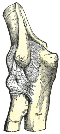
The humerus is a long bone in the arm that runs from the shoulder to the elbow. It connects the scapula and the two bones of the lower arm, the radius and ulna, and consists of three sections. The humeral upper extremity consists of a rounded head, a narrow neck, and two short processes. The body is cylindrical in its upper portion, and more prismatic below. The lower extremity consists of 2 epicondyles, 2 processes, and 3 fossae. As well as its true anatomical neck, the constriction below the greater and lesser tubercles of the humerus is referred to as its surgical neck due to its tendency to fracture, thus often becoming the focus of surgeons.
The forearm is the region of the upper limb between the elbow and the wrist. The term forearm is used in anatomy to distinguish it from the arm, a word which is used to describe the entire appendage of the upper limb, but which in anatomy, technically, means only the region of the upper arm, whereas the lower "arm" is called the forearm. It is homologous to the region of the leg that lies between the knee and the ankle joints, the crus.

In human anatomy, the ulnar nerve is a nerve that runs near the ulna bone. The ulnar collateral ligament of elbow joint is in relation with the ulnar nerve. The nerve is the largest in the human body unprotected by muscle or bone, so injury is common. This nerve is directly connected to the little finger, and the adjacent half of the ring finger, innervating the palmar aspect of these fingers, including both front and back of the tips, perhaps as far back as the fingernail beds.

The radius or radial bone is one of the two large bones of the forearm, the other being the ulna. It extends from the lateral side of the elbow to the thumb side of the wrist and runs parallel to the ulna. The ulna is longer than the radius, but the radius is thicker. The radius is a long bone, prism-shaped and slightly curved longitudinally.

The ulnar collateral ligament (UCL) or internal lateral ligament is a thick triangular ligament at the medial aspect of the elbow uniting the distal aspect of the humerus to the proximal aspect of the ulna.

The olecranon, is a large, thick, curved bony process on the proximal, posterior end of the ulna. It forms the protruding part of the elbow and is opposite to the cubital fossa or elbow pit. The olecranon serves as a lever for the extensor muscles that straighten the elbow joint.

The lateral epicondyle of the humerus is a large, tuberculated eminence, curved a little forward, and giving attachment to the radial collateral ligament of the elbow joint, and to a tendon common to the origin of the supinator and some of the extensor muscles. Specifically, these extensor muscles include the anconeus muscle, the supinator, extensor carpi radialis brevis, extensor digitorum, extensor digiti minimi, and extensor carpi ulnaris. In birds, where the arm is somewhat rotated compared to other tetrapods, it is termed dorsal epicondyle of the humerus. In comparative anatomy, the term ectepicondyle is sometimes used.
The pronator teres is a muscle that, along with the pronator quadratus, serves to pronate the forearm. It has two origins, at the medial humeral supracondylar ridge and the ulnar tuberosity, and inserts near the middle of the radius.
In human anatomy, the supinator is a broad muscle in the posterior compartment of the forearm, curved around the upper third of the radius. Its function is to supinate the forearm.

The posterior interosseous artery is an artery of the forearm. It is a branch of the common interosseous artery, which is a branch of the ulnar artery.

The annular ligament is a strong band of fibers that encircles the head of the radius, and retains it in contact with the radial notch of the ulna.

The medial epicondyle of the humerus is an epicondyle of the humerus bone of the upper arm in humans. It is larger and more prominent than the lateral epicondyle and is directed slightly more posteriorly in the anatomical position. In birds, where the arm is somewhat rotated compared to other tetrapods, it is called the ventral epicondyle of the humerus. In comparative anatomy, the more neutral term entepicondyle is used.

The anterior ligament of the elbow is a broad and thin fibrous layer covering the anterior surface of the joint.

The fascial compartments of arm refers to the specific anatomical term of the compartments within the upper segment of the upper limb of the body. The upper limb is divided into two segments, the arm and the forearm. Each of these segments is further divided into two compartments which are formed by deep fascia – tough connective tissue septa (walls). Each compartment encloses specific muscles and nerves.
The coronoid process of the ulna is a triangular process projecting forward from the anterior proximal portion of the ulna.
The humeroradial joint is the joint between the head of the radius and the capitulum of the humerus, is a limited ball-and-socket joint, hinge type of synovial joint.
The humeroulnar joint is part of the elbow-joint. It is composed of two bones, the humerus and ulna, and is the junction between the trochlear notch of ulna and the trochlea of humerus. It is classified as a simple hinge-joint, which allows for movements of flexion, extension and circumduction. Owing to the obliquity of the trochlea of the humerus, this movement does not take place in the antero-posterior plane of the body of the humerus.

The elbow is the region between the upper arm and the forearm that surrounds the elbow joint. The elbow includes prominent landmarks such as the olecranon, the cubital fossa, and the lateral and the medial epicondyles of the humerus. The elbow joint is a hinge joint between the arm and the forearm; more specifically between the humerus in the upper arm and the radius and ulna in the forearm which allows the forearm and hand to be moved towards and away from the body. The term elbow is specifically used for humans and other primates, and in other vertebrates it is not used. In those cases, forelimb plus joint is used.

