Palmar plate

Last updated
Palmar plate
Metacarpophalangeal joint illustration.svg
MCP joint in extension and in flexion, palmar plate in grey
Details
Identifiers
Latin ligamenta palmaria
MeSH D053401
TA98 A03.5.11.603
TA2 1844
FMA 71410
Anatomical terminology

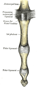
In the human hand, palmar or volar plates (also referred to as palmar or volar ligaments ) [1] are found in the metacarpophalangeal (MCP) and interphalangeal (IP) joints, where they reinforce the joint capsules, enhance joint stability, and limit hyperextension. The plates of the MCP and IP joints are structurally and functionally similar, except that in the MCP joints they are interconnected by a deep transverse ligament. In the MCP joints, they also indirectly provide stability to the longitudinal palmar arches of the hand. [2] [3] The volar plate of the thumb MCP joint has a transverse longitudinal rectangular shape, shorter than those in the fingers. [4]

Contents

Structure

This fibrocartilaginous structure is attached to the volar base of the phalanx distal to the joint. From there, it forms a palmar continuation of the articular surface of the phalanx bone and its inner surface thus adds to the articular surface during extension. [2]

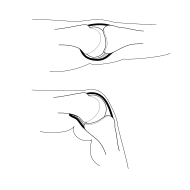
In its proximal end, the volar plate becomes membranous and blends with the volar capsule which is attached to the head of the metacarpal bone. During flexion, the plate glides proximally down the volar surface of the metacarpal head. Its flexible attachment to the phalanx bone not only prevents it from restricting joint movements, but also prevents the long flexor tendons from being pinched in the joint. [2] Flexion of the proximal phalanx is facilitated by the shape of the proximal edge, known as the volar recess, [5] but this diaphanous end of the volar plate is also the part of the metacarpophalangeal joint that is most susceptible to injury during dislocations. [6]

Due to its fibrocartilaginous composition, the plate is thus able to (1) resist tensile stresses while (2) restricting hyperextension and compression and (3) protecting the volar articular surface. [2]

Function

The palmar plate moves in three phases during joint flexion. First, it slides back toward the hand. Next, it is lifted away from the proximal phalanx by the A3 pulley. Last, a lip on the middle phalanx rolls into a recess on the plate. If the A3 pulley is not intact, the normal three phases of motion do not occur and instead the plate crumples. [7]

Metacarpophalangeal joints

In the MCP joints, the four volar plates of the fingers and the capsules within which they lie are blended with and interconnected by the deep transverse metacarpal ligament which ties the metacarpal heads together. Dorsal to this ligament on each side of the metacarpal heads are sagittal bands that connect the volar plates to the tendon of the extensor digitorum and to the extensor expansion. These bands help stabilise the volar plates over the metacarpal heads. [2]

In contrast to the volar plates of the MCP joints of the fingers, the volar plate of the thumb MCP joint is a thick structure firmly attached to the base of the proximal phalanx. It forms the bottom of a two-sided box, the sides of which are made up of the collateral ligaments. [8]

Additional images

See also

Notes

  1. In older literature they are also known as the glenoid ligaments of Cruveilhier or vaginal ligaments.
  2. 1 2 3 4 5 Austin 2005 , pp. 321–22 (MCP joints)
  3. Austin 2005 , p. 324 (IP joints)
  4. Schmidt & Lanz 2003 , p. 111
  5. Berger & Weiss 2004 , p. 175
  6. Gammons 2008 , Functional Anatomy
  7. Saito, S.; Suzuki, Y. (2011). "Biomechanics of the Volar Plate of the Proximal Interphalangeal Joint: A Dynamic Ultrasonographic Study". The Journal of Hand Surgery. 36 (2): 265–271. doi:10.1016/j.jhsa.2010.10.034. hdl: 2433/159397 . PMID   21276889.
  8. Doyle & Botte 2003 , pp. 540–41

Related Research Articles

<span class="mw-page-title-main">Carpal bones</span> Eight small bones that make up the wrist (or carpus) that connects the hand to the forearm

The carpal bones are the eight small bones that make up the wrist (carpus) that connects the hand to the forearm. The term "carpus" and "carpal" is derived from the Latin carpus and the Greek καρπός (karpós), meaning "wrist". In human anatomy, the main role of the carpal bones is to articulate with the radial and ulnar heads to form a highly mobile condyloid joint, to provide attachments for thenar and hypothenar muscles, and to form part of the rigid carpal tunnel which allows the median nerve and tendons of the anterior forearm muscles to be transmitted to the hand and fingers.

<span class="mw-page-title-main">Wrist</span> Part of the arm between the lower arm and the hand

In human anatomy, the wrist is variously defined as (1) the carpus or carpal bones, the complex of eight bones forming the proximal skeletal segment of the hand; (2) the wrist joint or radiocarpal joint, the joint between the radius and the carpus and; (3) the anatomical region surrounding the carpus including the distal parts of the bones of the forearm and the proximal parts of the metacarpus or five metacarpal bones and the series of joints between these bones, thus referred to as wrist joints. This region also includes the carpal tunnel, the anatomical snuff box, bracelet lines, the flexor retinaculum, and the extensor retinaculum.

<span class="mw-page-title-main">Metacarpal bones</span> Bones of hand

In human anatomy, the metacarpal bones or metacarpus, also known as the "palm bones", are the appendicular bones that form the intermediate part of the hand between the phalanges (fingers) and the carpal bones, which articulate with the forearm. The metacarpal bones are homologous to the metatarsal bones in the foot.

<span class="mw-page-title-main">Phalanx bone</span> Digital bone in the hands and feet of most vertebrates

The phalanges are digital bones in the hands and feet of most vertebrates. In primates, the thumbs and big toes have two phalanges while the other digits have three phalanges. The phalanges are classed as long bones.

<span class="mw-page-title-main">Extensor digitorum muscle</span> Muscle of the posterior forearm

The extensor digitorum muscle is a muscle of the posterior forearm present in humans and other animals. It extends the medial four digits of the hand. Extensor digitorum is innervated by the posterior interosseous nerve, which is a branch of the radial nerve.

In human anatomy, the extensor pollicis longus muscle (EPL) is a skeletal muscle located dorsally on the forearm. It is much larger than the extensor pollicis brevis, the origin of which it partly covers and acts to stretch the thumb together with this muscle.

<span class="mw-page-title-main">Palmar interossei muscles</span> Muscles between the metacarpals

In human anatomy, the palmar or volar interossei are four muscles, one on the thumb that is occasionally missing, and three small, unipennate, central muscles in the hand that lie between the metacarpal bones and are attached to the index, ring, and little fingers. They are smaller than the dorsal interossei of the hand.

<span class="mw-page-title-main">Metacarpophalangeal joint</span> Bodily joint at the base of each finger

The metacarpophalangeal joints (MCP) are situated between the metacarpal bones and the proximal phalanges of the fingers. These joints are of the condyloid kind, formed by the reception of the rounded heads of the metacarpal bones into shallow cavities on the proximal ends of the proximal phalanges. Being condyloid, they allow the movements of flexion, extension, abduction, adduction and circumduction at the joint.

<span class="mw-page-title-main">Carpometacarpal joint</span>

The carpometacarpal (CMC) joints are five joints in the wrist that articulate the distal row of carpal bones and the proximal bases of the five metacarpal bones.

<span class="mw-page-title-main">Dorsal interossei of the hand</span> Muscles between the metacarpals

In human anatomy, the dorsal interossei (DI) are four muscles in the back of the hand that act to abduct (spread) the index, middle, and ring fingers away from hand's midline and assist in flexion at the metacarpophalangeal joints and extension at the interphalangeal joints of the index, middle and ring fingers.

<span class="mw-page-title-main">Abductor digiti minimi muscle of hand</span> Muscle in the hypothenar compartment

In human anatomy, the abductor digiti minimi is a skeletal muscle situated on the ulnar border of the palm of the hand. It forms the ulnar border of the palm and its spindle-like shape defines the hypothenar eminence of the palm together with the skin, connective tissue, and fat surrounding it. Its main function is to pull the little finger away from the other fingers.

<span class="mw-page-title-main">Interphalangeal joints of the hand</span> Hinge joints between finger phalanges

The interphalangeal joints of the hand are the hinge joints between the phalanges of the fingers that provide flexion towards the palm of the hand.

<span class="mw-page-title-main">First metacarpal bone</span>

The first metacarpal bone or the metacarpal bone of the thumb is the first bone proximal to the thumb. It is connected to the trapezium of the carpus at the first carpometacarpal joint and to the proximal thumb phalanx at the first metacarpophalangeal joint.

<span class="mw-page-title-main">Intercarpal joints</span> Joints of the carpal bones of the wrist

The intercarpal joints can be subdivided into three sets of joints : Those of the proximal row of carpal bones, those of the distal row of carpal bones, and those of the two rows with each other.

<span class="mw-page-title-main">Annular ligaments of fingers</span>

In human anatomy, the annular ligaments of the fingers, often referred to as A pulleys, are the annular part of the fibrous sheathes of the fingers. Four or five such annular pulleys, together with three cruciate pulleys, form a fibro-osseous tunnel on the palmar aspect of the hand through which passes the deep and superficial flexor tendons. The annular and cruciate ligaments serve to govern the flexor mechanism of the hand and wrist, providing critical constraints to the flexor tendons to prevent bowstringing upon contraction and excursion of extrinsic flexor musculo-tendinous units.

<span class="mw-page-title-main">Collateral ligaments of metacarpophalangeal joints</span>

In human anatomy, the radial (RCL) and ulnar (UCL) collateral ligaments of the metacarpophalangeal joints (MCP) of the hand are the primary stabilisers of the MCP joints. A collateral ligament flanks each MCP joint - one on either side. Each attaches proximally at the head of the metacarpal bone, and distally at the base of the phalynx. Each extends obliquely in a palmar direction from its proximal attachment to its distal attachment. The collateral ligaments allow spreading our the fingers with an open hand but not with the hand closed into a fist.

In the human foot, the plantar or volar plates are fibrocartilaginous structures found in the metatarsophalangeal (MTP) and interphalangeal (IP) joints. The anatomy and composition of the plantar plates are similar to the palmar plates in the metacarpophalangeal (MCP) and interphalangeal joints in the hand; the proximal origin is thin but the distal insertion is stout. Due to the weight-bearing nature of the human foot, the plantar plates are exposed to extension forces not present in the human hand.

<span class="mw-page-title-main">Ulnar claw</span> Deformity of the hand that develops due to ulnar nerve damage

An ulnar claw, also known as claw hand, is a deformity or an abnormal attitude of the hand that develops due to ulnar nerve damage causing paralysis of the lumbricals. A claw hand presents with a hyperextension at the metacarpophalangeal joints and flexion at the proximal and distal interphalangeal joints of the 4th and 5th fingers. The patients with this condition can make a full fist but when they extend their fingers, the hand posture is referred to as claw hand. The ring- and little finger can usually not fully extend at the proximal interphalangeal joint (PIP).

<span class="mw-page-title-main">Hand</span> Extremity at the end of an arm or forelimb

A hand is a prehensile, multi-fingered appendage located at the end of the forearm or forelimb of primates such as humans, chimpanzees, monkeys, and lemurs. A few other vertebrates such as the koala are often described as having "hands" instead of paws on their front limbs. The raccoon is usually described as having "hands" though opposable thumbs are lacking.

<span class="mw-page-title-main">Extrinsic extensor muscles of the hand</span>

The extrinsic extensor muscles of the hand are located in the back of the forearm and have long tendons connecting them to bones in the hand, where they exert their action. Extrinsic denotes their location outside the hand. Extensor denotes their action which is to extend, or open flat, joints in the hand. They include the extensor carpi radialis longus (ECRL), extensor carpi radialis brevis (ECRB), extensor digitorum (ED), extensor digiti minimi (EDM), extensor carpi ulnaris (ECU), abductor pollicis longus (APL), extensor pollicis brevis (EPB), extensor pollicis longus (EPL), and extensor indicis (EI).

References