Related Research Articles

The foot is an anatomical structure found in many vertebrates. It is the terminal portion of a limb which bears weight and allows locomotion. In many animals with feet, the foot is a separate organ at the terminal part of the leg made up of one or more segments or bones, generally including claws or nails.

The human leg, in the general word sense, is the entire lower limb of the human body, including the foot, thigh and even the hip or gluteal region. However, the definition in human anatomy refers only to the section of the lower limb extending from the knee to the ankle, also known as the crus or, especially in non-technical use, the shank. Legs are used for standing, and all forms of locomotion including recreational such as dancing, and constitute a significant portion of a person's mass. Female legs generally have greater hip anteversion and tibiofemoral angles, but shorter femur and tibial lengths than those in males.

The extensor digitorum muscle is a muscle of the posterior forearm present in humans and other animals. It extends the medial four digits of the hand. Extensor digitorum is innervated by the posterior interosseous nerve, which is a branch of the radial nerve.
In human anatomy, the dorsal interossei of the foot are four muscles situated between the metatarsal bones.

The extensor digitorum longus is a pennate muscle, situated at the lateral part of the front of the leg.

The Adductor hallucis arises by two heads—oblique and transverse and is responsible for adducting the big toe. It has two heads, both are innervated by the lateral plantar nerve.

Flexor hallucis brevis muscle is a muscle of the foot that flexes the big toe.

In human anatomy, plantar interossei muscles are three muscles located between the metatarsal bones in the foot.

The lumbricals are four small skeletal muscles, accessory to the tendons of the flexor digitorum longus muscle. They are numbered from the medial side of the foot.
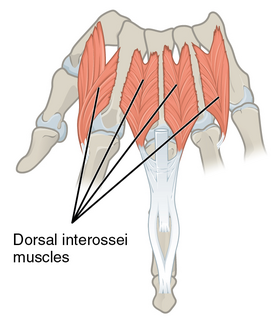
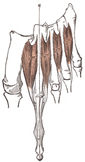
In human anatomy, the dorsal interossei (DI) are four muscles in the back of the hand that act to abduct (spread) the index, middle, and ring fingers away from hand's midline and assist in flexion at the metacarpophalangeal joints and extension at the interphalangeal joints of the index, middle and ring fingers.

The interphalangeal joints of the hand are the hinge joints between the phalanges of the fingers that provide flexion towards the palm of the hand.

The sole is the bottom of the foot.

The metatarsophalangeal joints are the joints between the metatarsal bones of the foot and the proximal bones of the toes. They are condyloid joints, meaning that an elliptical or rounded surface comes close to a shallow cavity.

The arches of the foot, formed by the tarsal and metatarsal bones, strengthened by ligaments and tendons, allow the foot to support the weight of the body in the erect posture with the least weight.
The transverse metatarsal ligament is a narrow band which runs across and connects together the heads of all the metatarsal bones. It is blended anteriorly with the plantar (glenoid) ligaments of the metatarsophalangeal articulations.

The interphalangeal joints of the foot are between the phalanx bones of the toes in the feet.

The fourth metatarsal bone is a long bone in the foot. It is smaller in size than the third metatarsal bone and is the third longest of the five metatarsal bones. The fourth metatarsal is analogous to the fourth metacarpal bone in the hand

The third metatarsal bone is a long bone in the foot. It is the second longest metatarsal. The longest being the second metatarsal. The third metatarsal is analogous to the third metacarpal bone in the hand

The second metatarsal bone is a long bone in the foot. It is the longest of the metatarsal bones, being prolonged backward and held firmly into the recess formed by the three cuneiform bones. The second metatarsal forms joints with the second proximal phalanx through the metatarsophalangeal joint, the cuneiform bones, third metatarsal and occasionally the first metatarsal bone.

The following outline is provided as an overview of and topical guide to human anatomy:
References
- Canale, S. Terry; Beaty, James H.; Ishikawa, Susan N. (2008). "Hammer Toe Correction". Procedures Consult (Elsevier). Archived from the original on 2010-07-26. Retrieved 25 November 2010.
- Deland, JT; Lee, KT; Sobel, M; DiCarlo, EF (August 1995). "Anatomy of the plantar plate and its attachments in the lesser metatarsal phalangeal joint". Foot Ankle Int. 16 (8): 480–6. doi:10.1177/107110079501600804. PMID 8520660. S2CID 24841493.
- Johnston, RB 3rd; Smith, J; Daniels, T (May 1994). "The plantar plate of the lesser toes: an anatomical study in human cadavers". Foot Ankle Int. 15 (5): 276–82. doi:10.1177/107110079401500508. PMID 7951967. S2CID 41157698.
- Mohana-Borges, Aurea V. R.; Theumann, Nicolas H.; Pfirrmann, Christian W. A.; Chung, Christine B.; Resnick, Donald L.; Trudell, Debra J. (April 2003). "Lesser Metatarsophalangeal Joints: Standard MR Imaging, MR Arthrography, and MR Bursography—Initial Results in 48 Cadaveric Joints". Radiology. 227 (1): 175–182. doi:10.1148/radiol.2271020283. PMID 12668744.
- Stainsby, G D (January 1997). "Pathological anatomy and dynamic effect of the displaced plantar plate and the importance of the integrity of the plantar plate-deep transverse metatarsal ligament tie-bar". Annals of the Royal College of Surgeons of England. 79 (1): 58–68. PMC 2502622 . PMID 9038498.