Plantar fascia

Last updated
Plantar fascia
1124 Intrinsic Muscles of the Foot b.png
Muscles of the sole of the foot. First layer (closest to the skin on the sole of the foot). Plantar aponeurosis visible at top center.
Details
Identifiers
Latin aponeurosis plantaris
TA98 A04.7.03.031
TA2 2718
FMA 45171
Anatomical terminology

The plantar fascia or plantar aponeurosis [1] is the thick connective tissue aponeurosis which supports the arch on the bottom (plantar side) of the foot. Recent studies suggest that the plantar fascia is actually an aponeurosis rather than true fascia.[ citation needed ] It runs from the tuberosity of the calcaneus (heel bone) forward to the heads of the metatarsal bones (the bone between each toe and the bones of the mid-foot).

Contents

Anatomy

Anatomical diagrams illustrating the components of the plantar fascia. PF-PlantarDesign.jpg
Anatomical diagrams illustrating the components of the plantar fascia.
Dissection of the plantar aponeurosis:
LP, lateral part; CP, central part; MP, medial part; L, length; W, width. Plantar aponeurosis - axial view.png
Dissection of the plantar aponeurosis:
LP, lateral part; CP, central part; MP, medial part; L, length; W, width.
Five central part plantar aponeurosis bundles. Plantar aponeurosis - central.png
Five central part plantar aponeurosis bundles.

The plantar fascia is the thick central portion of the fascia investing the plantar muscles. It extends between the medial process of the tuber calcanei [1] and the proximal phalanges of[ citation needed ] the toes. It provides some attachment to the flexor muscles of the toes. [1]

Distally, the plantar fascia becomes continuous with the fibrous sheats enveloping the flexor tendons passing to the toes. At the anterior extremity of the sole - inferior to the heads of the metatarsal bones - the plantar aponeurosis forms the superficial transverse metatarsal ligament. [2]

Structure

The plantar fascia is made up of predominantly longitudinally oriented collagen fibers. There are three distinct structural components: the medial component, the central component (plantar aponeurosis), and the lateral component (see diagram at right). The central component is the largest and most prominent.

Development

In younger people the plantar fascia is also intimately related to the Achilles tendon, with a continuous fascial connection between the two from the distal aspect of the Achilles to the origin of the plantar fascia at the calcaneal tubercle. However, the continuity of this connection decreases with age to a point that in the elderly there are few, if any, connecting fibers. There are also distinct attachments of the plantar fascia and the Achilles tendon to the calcaneus so the two do not directly contact each other. Nevertheless, there is an indirect relationship whereby if the toes are dorsiflexed, the plantar fascia tightens via the windlass mechanism. If a tensile force is then generated in the Achilles tendon it will increase tensile strain in the plantar fascia. Clinically, this relationship has been used as a basis for treatment for plantar fasciitis, with stretches and night stretch splinting being applied to the gastrocnemius/soleus muscle unit.

Function

The effect of dorsiflexing the toes on arch height (A). The windlass mechanism (B). Plantar Bewegung.gif
The effect of dorsiflexing the toes on arch height (A). The windlass mechanism (B).

The plantar fascia contributes to support of arch of the foot by acting as a tie-rod, where it undergoes tension when the foot bears weight. One biomechanical model estimated it carries as much as 14% of the total load of the foot. In an experiment using cadavers, it was found that failure of the plantar fascia averaged at loads of 1189 ± 244 newtons [3] (121 ± 24 kgf or 267 ± 55 lbf). Failure most often occurred at the proximal attachment to the calcaneus, which is consistent with the usual location of symptoms (i.e. in plantar fasciitis). Complete rupture or surgical release of the plantar fascia leads to a decrease in arch stiffness and a significant collapse of the longitudinal arch of the foot. By modeling it was predicted such conditions would result in a 17% increase in vertical displacement and a 15% increase in horizontal elongation of the foot when it was loaded at 683 newtons (154 lbf). [4] Surgical release also significantly increases both stress in the plantar ligaments and plantar pressures under the metatarsal heads. Although most of the figures mentioned above are from either cadaver studies or investigations using models, they highlight the relatively large load the plantar fascia is subjected to while contributing to the structural integrity of the foot.

Gait

The plantar fascia also has an important role in dynamic function during gait. It was found the plantar fascia continuously elongated during the contact phase of gait. It went through rapid elongation before and immediately after mid-stance, reaching a maximum of 9% to 12% elongation between mid-stance and toe-off. [5] During this phase the plantar fascia behaves like a spring, which may assist in conserving energy. In addition, the plantar fascia has a critical role in normal mechanical function of the foot, contributing to the "windlass mechanism". When the toes are dorsiflexed in the propulsive phase of gait, the plantar fascia becomes tense, resulting in elevation of the longitudinal arch and shortening of the foot (see 3A). One can liken this mechanism to a cable being wound around the drum of a windlass (see 3B); the plantar fascia being the cable, the metatarsal head the drum, and the handle, the proximal phalanx.

Clinical significance

Plantar fasciitis

Other

Additional images

See also

Related Research Articles

<span class="mw-page-title-main">Foot</span> Anatomical structure found in vertebrates

The foot is an anatomical structure found in many vertebrates. It is the terminal portion of a limb which bears weight and allows locomotion. In many animals with feet, the foot is a separate organ at the terminal part of the leg made up of one or more segments or bones, generally including claws and or nails.

<span class="mw-page-title-main">Human leg</span> Lower extremity or limb of the human body (foot, lower leg, thigh and hip)

The human leg is the entire lower limb of the human body, including the foot, thigh or sometimes even the hip or buttock region. The major bones of the leg are the femur, tibia, and adjacent fibula. The thigh is between the hip and knee, while the calf (rear) and shin (front) are between the knee and foot.

<span class="mw-page-title-main">Heel</span> Part of the foot in the back

The heel is the prominence at the posterior end of the foot. It is based on the projection of one bone, the calcaneus or heel bone, behind the articulation of the bones of the lower leg.

<span class="mw-page-title-main">Fibularis longus</span> Superficial muscle in the lateral compartment of the leg

In human anatomy, the fibularis longus is a superficial muscle in the lateral compartment of the leg. It acts to tilt the sole of the foot away from the midline of the body (eversion) and to extend the foot downward away from the body at the ankle.

<span class="mw-page-title-main">Achilles tendon</span> Tendon at the back of the lower leg

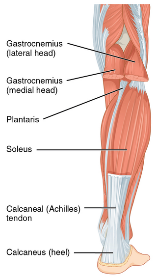
The Achilles tendon or heel cord, also known as the calcaneal tendon, is a tendon at the back of the lower leg, and is the thickest in the human body. It serves to attach the plantaris, gastrocnemius (calf) and soleus muscles to the calcaneus (heel) bone. These muscles, acting via the tendon, cause plantar flexion of the foot at the ankle joint, and flexion at the knee.

<span class="mw-page-title-main">Calcaneus</span> Bone of the tarsus of the foot

In humans and many other primates, the calcaneus or heel bone is a bone of the tarsus of the foot which constitutes the heel. In some other animals, it is the point of the hock.

<span class="mw-page-title-main">Pes cavus</span> Medical condition

Pes cavus, also known as high arch, is a human foot type in which the sole of the foot is distinctly hollow when bearing weight. That is, there is a fixed plantar flexion of the foot. A high arch is the opposite of a flat foot and is somewhat less common.

<span class="mw-page-title-main">Plantar fasciitis</span> Connective tissue disorder of the heel

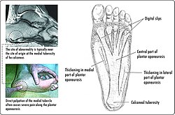
Plantar fasciitis or plantar heel pain is a disorder of the plantar fascia, which is the connective tissue which supports the arch of the foot. It results in pain in the heel and bottom of the foot that is usually most severe with the first steps of the day or following a period of rest. Pain is also frequently brought on by bending the foot and toes up towards the shin. The pain typically comes on gradually, and it affects both feet in about one-third of cases.

<span class="mw-page-title-main">Flat feet</span> Deformity in which the foot arches contact the ground

Flat feet is a postural deformity in which the arches of the foot collapse, with the entire sole of the foot coming into complete or near-complete contact with the ground. Sometimes children are born with flat feet (congenital).

<span class="mw-page-title-main">Tarsus (skeleton)</span> Bones of the foot

In the human body, the tarsus is a cluster of seven articulating bones in each foot situated between the lower end of the tibia and the fibula of the lower leg and the metatarsus. It is made up of the midfoot and hindfoot.

<span class="mw-page-title-main">Dorsal interossei of the foot</span> Four muscles situated between the metatarsal bones

In human anatomy, the dorsal interossei of the foot are four muscles situated between the metatarsal bones.

<span class="mw-page-title-main">Flexor hallucis brevis muscle</span> Muscle in sole of the foot that leads to the big toe

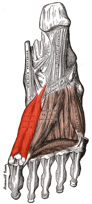
Flexor hallucis brevis muscle is a muscle of the foot that flexes the big toe.

<span class="mw-page-title-main">Calcaneal spur</span> Medical condition of the heel

A calcaneal spur is a bony outgrowth from the calcaneal tuberosity. Calcaneal spurs are typically detected by x-ray examination. It is a form of exostosis.

<span class="mw-page-title-main">Sole (foot)</span> Bottom part of foot

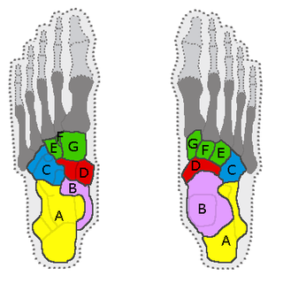
The sole is the bottom of the foot.

<span class="mw-page-title-main">Arches of the foot</span> Load-bearing curves in the tarsal and metatarsal bones of the feet

The arches of the foot, formed by the tarsal and metatarsal bones, strengthened by ligaments and tendons, allow the foot to support the weight of the body in the erect posture with the least weight.

<span class="mw-page-title-main">Fifth metatarsal bone</span>

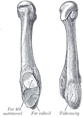
The fifth metatarsal bone is a long bone in the foot, and is palpable along the distal outer edges of the feet. It is the second smallest of the five metatarsal bones. The fifth metatarsal is analogous to the fifth metacarpal bone in the hand.

The Ponseti method is a manipulative technique that corrects congenital clubfoot without invasive surgery. It was developed by Ignacio V. Ponseti of the University of Iowa Hospitals and Clinics, USA in the 1950s, and was repopularized in 2000 by John Herzenberg in the US and Europe and in Africa by NHS surgeon Steve Mannion. It is a standard treatment for clubfoot.

In the human foot, the plantar or volar plates are fibrocartilaginous structures found in the metatarsophalangeal (MTP) and interphalangeal (IP) joints. The anatomy and composition of the plantar plates are similar to the palmar plates in the metacarpophalangeal (MCP) and interphalangeal joints in the hand; the proximal origin is thin but the distal insertion is stout. Due to the weight-bearing nature of the human foot, the plantar plates are exposed to extension forces not present in the human hand.

<span class="mw-page-title-main">Locomotor effects of shoes</span>

Locomotor effects of shoes are the way in which the physical characteristics or components of shoes influence the locomotion neuromechanics of a person. Depending on the characteristics of the shoes, the effects are various, ranging from alteration in balance and posture, muscle activity of different muscles as measured by electromyography (EMG), and the impact force. There are many different types of shoes that exist, such as running, walking, loafers, high heels, sandals, slippers, work boots, dress shoes, and many more. However, a typical shoe will be composed of an insole, midsole, outsole, and heels, if any. In an unshod condition, where one is without any shoes, the locomotor effects are primarily observed in the heel strike patterns and resulting impact forces generated on the ground.

<span class="mw-page-title-main">Plantar fascial rupture</span> Medical condition

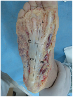
A plantar fascial rupture, is a painful tear in the plantar fascia. The plantar fascia is a connective tissue that spans across the bottom of the foot. The condition plantar fasciitis may increase the likelihood of rupture. A plantar fascial rupture may be mistaken for plantar fasciitis or even a calcaneal fracture. To allow for proper diagnosis, an MRI is often needed.

References

  1. 1 2 3 "aponeurosis plantaris". TheFreeDictionary.com. Retrieved 2023-06-10.
  2. Moore, Keith L.; Dalley, Arthur F.; Agur, Anne M. R. (2017). Essential Clinical Anatomy. Lippincott Williams & Wilkins. p. 769. ISBN   978-1496347213.
  3. H. B. Kitaoka; Z. P. Luo; E. S. Growney; L. J. Berglund; K. N. An (October 1994). "Material properties of the plantar aponeurosis". Foot & Ankle International. 15 (10): 557–560. doi:10.1177/107110079401501007. PMID   7834064. S2CID   45264072.
  4. G. A. Arangio, C. Chen and W. Kim (June 1997). "Effect of cutting the plantar fascia on mechanical properties of the foot". Clinical Orthopaedics and Related Research. 339 (339): 227–231. doi:10.1097/00003086-199706000-00031. PMID   9186224. S2CID   19194412.
  5. Amit Gefen (March 2003). "The in vivo elastic properties of the plantar fascia during the contact phase of walking". Foot & Ankle International. 24 (3): 238–244. doi:10.1177/107110070302400307. PMID   12793487. S2CID   2829705.
  6. "Plantar Fascial Tears". American Foot & Leg Specialists. 2016-10-17. Retrieved 2018-04-23.