The cause is unknown.[3] The underlying mechanism is believed to involve an outpouching of the synovial membrane.[4] Diagnosis is typically based on examination. The ability to shine light through the bump or any past decrease in size supports the diagnosis of the bump as a ganglion cyst.[4] Ganglion cysts are usually obvious upon observation. Medical imaging may be considered on infrequent occasions to rule out another diagnosis.[3][4]
Treatment is not necessary. Options for treatment include needle aspiration or surgery.[3] About half the time, they resolve on their own.[4] About three per 10,000 people develop a ganglion cyst of the wrist or hand a year.[5]
Presentation
The average size of these cysts is 2 centimetres (0.79in), but excised cysts of more than 5 centimetres (2.0in) have been reported.[6] The size of the cyst may vary over time. Between 50 and 70% of all masses on the hand and wrist are ganglion cysts.[7][8][9]
Wrist
They commonly are found near the wrist joint, especially at the scapholunate area.[10]
In a 2007 study of patients in Glasgow whose foot lumps were removed surgically, 39 of 101 cases were ganglion cysts. The study replicated earlier findings that no ganglion cysts were found on the sole or heel. The authors wrote, "Although lumps in these areas may be ganglia, the surgeon should probably consider other diagnoses in the first instance." The researchers noted a preponderance of occurrence among females (85%) and that 11 of the other cases had been misdiagnosed as ganglion cysts before surgery.[14]
Lower extremity
Ganglion cysts are not limited to the hands and feet. They may occur near the knee, mostly within and near the anterior cruciate ligament,[9] but they may occur at the origins of the gastrocnemius tendon, and anteriorly on Hoffa's infrapatellar fat pad.[15] Most patients with a ganglion cyst of the knee present with both pain and a restricted range of motion, but these findings are variable, and some patients may have neither.[9]
Other
From their common origin at a joint or tendon, ganglion cysts may form in a wide range of locations. At the shoulder, they typically occur at the acromioclavicular joint or along the biceps tendon,[16] and are occasionally known to cause nerve compression or bone erosion.[17] Rarely, intraosseous ganglion cysts occur, sometimes in combination with a cyst in the overlying soft tissue.[6][18] It is possible for a cyst to be considerably displaced from the joint. In one extreme case, a ganglion cyst was observed to propagate extensively via the conduit of the common peroneal nerve sheath to a location in the thigh; in such cases surgery to the proximal joint to remove the articular connection may remove the need for a riskier, more extensive surgery in the neural tissue of the thigh.[19] The cysts may intrude into the spine, which may cause pain and dysesthesia in distant extremities.[20]
Cystic adventitial disease, in which a cyst occurs within the popliteal artery near the knee, has been proposed recently to occur by an articular mechanism, with a conduit leading from the joint, similar to the development of ganglion cysts, that spreads within the peroneal nerve.[21] One uncommon location of the cysts are in the muscle tendons of the hand, such as the extensor digitorum superficialis.[22]
Cyst on right wrist
Cyst on dorsum of right foot
Cyst on a finger
Small cyst on right index finger
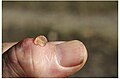
small cyst on thumb lanced with red-hot needle
Ganglion cyst on the palmar side of the left wrist
Causes
The exact cause is unknown.[9] The most commonly accepted probable cause of ganglion cysts is the herniation hypothesis, by which they are thought to occur as an out-pouching or distention of a weakened portion of a joint capsule or tendon sheath. This description is based on the observations that the cysts occur close to tendons and joints. The microscopic anatomy of the cyst resembles that of tenosynovial tissue. The fluid is similar in composition to synovial fluid. Dye injected into the joint frequently ends up in the cyst. Dye injected into the cyst rarely enters the joint, however, which has been attributed to the apparent formation of an effective and one-way "check valve", allowing fluid out of the joint, but not back in.[6]
Ganglion cyst of the hand with multiple cystic chambers containing glairy material - the walls are composed of bland fibrous tissue with no specialized lining.
Ganglion cysts are diagnosed easily, as they are visible and pliable to touch.
Ultrasonography (US) may be used to increase diagnostic confidence in clinically suspected lesions or to view smaller "occult" cysts as a cause of dorsal wrist pain with forceful extension.
Treatment
At least 33% resolve without treatment within six years, and 50% within 10 years.[24]
Surgical excision is the primary discretionary, elective treatment option for ganglion cysts. Alternatively, a hypodermic needle may be used to drain the fluid from the cyst (via aspiration).[25] The recurrence rate is about 50% following aspiration of a ganglion cyst.
Complications
Complications of treatment may include joint stiffness and scar formation.[25] Recurrence of the lesion is more common following excision of a volar ganglion cyst in the wrist. Incomplete excision that fails to include the stalk or pedicle also may lead to recurrence, as will failing to execute a layered closure of the incision.[26]
Prognosis
Recurrence rate is higher in aspirated cysts than in excised ones.[27] Ganglion cysts have been found to recur following surgery in 12%[28] to 41%[29] of patients.
A six-year outcome study of the treatment of ganglion cysts on the dorsal wrist compared excision, aspiration, and no treatment. Neither excision nor aspiration provided long-term benefit better than no treatment. Of the untreated ganglion cysts, 58% resolved spontaneously; the postsurgery recurrence rate in this study was 39%.[30] A similar study in 2003 of ganglion cysts occurring on the palmar surface of the wrist states: "At 2- and 5-year follow-up, regardless of treatment, no difference in symptoms was found, regardless of whether the palmar wrist ganglion was excised, aspirated, or left alone."[31]
Etymology
Being a misnomer that has persisted into modern times,[32] the ganglion cyst is unrelated to the neural ganglion or ganglion cell; its etymology traces back to the ancient Greek γάγγλιον, a 'knot' or 'swelling beneath the skin',[33] which extends to the neural masses by analogy. Generally, Hippocrates is credited with the description of these cysts.[6][34]
"Bible bump"
A historical method of treatment for a ganglion cyst was to strike the lump with a large, heavy book, causing the cyst to rupture and drain into the surrounding tissues. Historically, a Bible, usually the largest (or only) book in a household, was employed for this treatment. This practice led to the nickname of "Bible bumps" or "Gideon's disease" for the cysts.[2][35] This treatment risks injuring the person and thus is not recommended.[36][37]
↑"Ganglion Cyst". Medical Subject Headings (MeSH). National Library of Medicine. Archived from the original on March 10, 2016. Retrieved August 27, 2013.
↑Stretanski MF (2020). "32. Hand and Wrist Ganglia". In Frontera WR, Silver JK, Rizzo TD (eds.). Essentials of Physical Medicine and Rehabilitation (4thed.). Content Repository Only!. pp.169–173. ISBN978-0-323-54947-9. Retrieved 2020-10-24.
↑R. J. Spinner, etal. (2012-08-29). "Evidence to support that adventitial cysts, analogous to intraneural ganglion cysts, are also joint-connected". Clin. Anat. 26 (2): 267–81. doi:10.1002/ca.22152. PMID22933403. S2CID37986961.
↑Dias JJ, Dhukaram V, Kumar P (October 2007). "The natural history of untreated dorsal wrist ganglia and patient reported outcome 6 years after intervention". The Journal of Hand Surgery, European Volume. 32 (5): 502–8. doi:10.1016/J.JHSE.2007.05.007. PMID17950209. S2CID21853864.
↑Dias J, Buch K (Apr 2003). "Palmar wrist ganglion: does intervention improve outcome? A prospective study of the natural history and patient-reported treatment outcomes". J Hand Surg Br Vol. 28 (2): 172–6. doi:10.1016/s0266-7681(02)00365-0. PMID12631492. S2CID44865301.
↑J.C. Segen (1992). "Aneurysmal bone cyst". The Dictionary of Modern Medicine. CRC Press. ISBN978-1-85070-321-1. Archived from the original on 2017-09-10. like pyogenic granuloma and ganglion cyst, a misnomer that has withstood the sands of time and the dint of logic
This page is based on this Wikipedia article Text is available under the CC BY-SA 4.0 license; additional terms may apply. Images, videos and audio are available under their respective licenses.