The term "connective tissue" (in German, Bindegewebe) was introduced in 1830 by Johannes Peter Müller. The tissue was already recognized as a distinct class in the 18th century.[3][4]
Types
Connective tissue can be broadly classified into connective tissue proper (including loose connective tissue and dense connective tissue) and special connective tissue (including supportive connective tissue and fluid connective tissue).[5][6]
Connective tissue proper
Loose and dense connective tissue are distinguished by the ratio of ground substance to fibrous tissue. Loose connective tissue has much more ground substance and a relative lack of fibrous tissue, while the reverse is true of dense connective tissue.
Dense connective tissue is subdivided into dense regular and dense irregular connective tissue.[7] Dense regular connective tissue, found in structures such as tendons and ligaments, is characterized by collagen fibers arranged in an orderly parallel fashion, giving it tensile strength in one direction. Dense irregular connective tissue provides strength in multiple directions by its dense bundles of fibers arranged in all directions.[citation needed]
Special connective tissue
Special connective tissue consists of supportive connective tissue (comprising bone and cartilage) and fluid connective tissue (comprising blood and lymph).[1][8][9][10][11] Special connective tissue are a form of fascia, with blood and lymph being known as liquid fascia.[1][12][13]
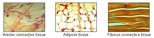
Other kinds of special connective tissues include fibrous, elastic, and lymphoid connective tissues.[14] Fibroareolar tissue is a mix of fibrous and areolar tissue.[15] Fibromuscular tissue is made up of fibrous tissue and muscular tissue.
New vascularised connective tissue that forms in the process of wound healing is termed granulation tissue.[16]
Ground substance is a clear, colorless, and viscous fluid containing glycosaminoglycans and proteoglycans allowing fixation of collagen fibers in intercellular spaces. Examples of non-fibrous connective tissue include adipose tissue (fat) and blood. Adipose tissue gives "mechanical cushioning" to the body, among other functions.[19][20] Although there is no dense collagen network in adipose tissue, groups of adipose cells are kept together by collagen fibers and collagen sheets in order to keep fat tissue under compression in place (for example, the sole of the foot). Both the ground substance and proteins create the matrix for connective tissue.
Type I collagen is present in many forms of connective tissue, and makes up about 25% of the total protein content of the mammalian body.[21]
Connective tissue has a wide variety of functions that depend on the types of cells and the different classes of fibers involved.
Loose and dense irregular connective tissue, formed mainly by fibroblasts and collagen fibers, have an important role in providing a medium for oxygen and nutrients to diffuse from capillaries to cells, and carbon dioxide and waste substances to diffuse from cells back into circulation. They also allow organs to resist stretching and tearing forces.[22]
Mesenchyme is a type of connective tissue found in the developing organs of an embryo that is capable of differentiation into all types of mature connective tissue.[23] Another type of relatively undifferentiated connective tissue is the mucous connective tissue known as Wharton's jelly, found inside the umbilical cord.[22] This tissue is no longer present after birth, leaving only scattered mesenchymal cells throughout the body.[24]
12345Biga, Lindsay M.; Dawson, Sierra; Harwell, Amy; Hopkins, Robin; Kaufmann, Joel; LeMaster, Mike; Matern, Philip; Morrison-Graham, Katie; Quick, Devon (2019), "4.3 Connective Tissue Supports and Protects", Anatomy & Physiology, OpenStax/Oregon State University, retrieved 16 April 2021
↑Biga, Lindsay M.; Dawson, Sierra; Harwell, Amy (26 September 2019). "4.1 Types of Tissues". Retrieved 30 July 2022.
↑Mathews, M. B. (1975). Connective Tissue, Macromolecular Structure Evolution. Springer-Verlag, Berlin and New York. link.
↑Aterman, K. (1981). "Connective tissue: An eclectic historical review with particular reference to the liver". The Histochemical Journal. 13 (3): 341–396. doi:10.1007/BF01005055. PMID7019165. S2CID22765625.
12345678910Ross, Michael H.; Pawlina, Wojciech (2011). Histology: a text and atlas; with correlated cell and molecular biology (6., internationaled.). Philadelphia, Pa. London: Lippincott Williams & Wilkins. pp.158–173. ISBN978-0781772006.
↑Young B, Woodford P, O'Dowd G (2013). Wheater's Functional Histology: A Text and Colour Atlas (6thed.). Elsevier. p.65. ISBN978-0702047473.
↑This article incorporates text available under the CC BY 4.0 license.Betts, J Gordon; Desaix, Peter; Johnson, Eddie; Johnson, Jody E; Korol, Oksana; Kruse, Dean; Poe, Brandon; Wise, James; Womble, Mark D; Young, Kelly A (26 June 2023). Anatomy & Physiology. Houston: OpenStax CNX. 4.3 Connective Tissue supports and protects. ISBN978-1-947172-04-3.
This page is based on this Wikipedia article Text is available under the CC BY-SA 4.0 license; additional terms may apply. Images, videos and audio are available under their respective licenses.