Serous membrane

Last updated
Serous membrane
Illu stomach2.jpg
Stomach. (Serosa is labeled at far right, and is colored yellow.)
Details
Precursor mesoderm
Identifiers
Latin tunica serosa
MeSH D012704
FMA 9581
Anatomical terminology
Serous membrane lines the pericardial cavity and reflects back to cover the heart--much the same way that an underinflated balloon would form two layers surrounding a fist. Serous Membrane.jpg
Serous membrane lines the pericardial cavity and reflects back to cover the heart—much the same way that an underinflated balloon would form two layers surrounding a fist.

The serous membrane (or serosa) is a smooth tissue membrane of mesothelium lining the contents and inner walls of body cavities, which secrete serous fluid to allow lubricated sliding movements between opposing surfaces. The serous membrane that covers internal organs is called visceral, while the one that covers the cavity wall is called parietal. For instance the parietal peritoneum is attached to the abdominal wall and the pelvic walls. [2] The visceral peritoneum is wrapped around the visceral organs. For the heart, the layers of the serous membrane are called parietal and visceral pericardium. For the lungs they are called parietal and visceral pleura. The visceral serosa of the uterus is called the perimetrium. The potential space between two opposing serosal surfaces is mostly empty except for the small amount of serous fluid. [3]

Contents

The Latin anatomical name is tunica serosa. Serous membranes line and enclose several body cavities, also known as serous cavities, where they secrete a lubricating fluid which reduces friction from movements. Serosa is entirely different from the adventitia, a connective tissue layer which binds together structures rather than reducing friction between them. The serous membrane covering the heart and lining the mediastinum is referred to as the pericardium, the serous membrane lining the thoracic cavity and surrounding the lungs is referred to as the pleura, and that lining the abdominopelvic cavity and the viscera is referred to as the peritoneum.

Structure

Serous membranes have two layers. The parietal layers of the membranes line the walls of the body cavity (pariet- refers to a cavity wall). The visceral layer of the membrane covers the organs (the viscera). Between the parietal and visceral layers is a very thin, fluid-filled serous space, or cavity. [4]

Visceral and parietal layers

Each serous membrane is composed of a secretory epithelial layer and a connective tissue layer underneath.

For the heart, the layers of the serous membrane are called the parietal pericardium, and the visceral pericardium (sometimes called the epicardium). Other parts of the body may also have specific names for these structures. For example, the serosa of the uterus is called the perimetrium.

Schematic diagram of an organ invaginating into a serous cavity Serous organ invagination.gif
Schematic diagram of an organ invaginating into a serous cavity

The pericardial cavity (surrounding the heart), pleural cavity (surrounding the lungs) and peritoneal cavity (surrounding most organs of the abdomen) are the three serous cavities within the human body. While serous membranes have a lubricative role to play in all three cavities, in the pleural cavity it has a greater role to play in the function of breathing.

The serous cavities are formed from the intraembryonic coelom and are basically an empty space within the body surrounded by serous membrane. Early in embryonic life visceral organs develop adjacent to a cavity and invaginate into the bag-like coelom. Therefore, each organ becomes surrounded by serous membrane - they do not lie within the serous cavity. The layer in contact with the organ is known as the visceral layer, while the parietal layer is in contact with the body wall.

Examples

In the human body, there are three serous cavities with associated serous membranes:

The two layers of serous membranes are named parietal and visceral. Between the two layers is a thin fluid filled space. [4] The fluid is produced by the serous membranes and stays between the two layers to reduce friction between the walls of the cavities and the internal organs when they move with respect to one another, such as when the lungs inflate or the heart beats. Such movement could otherwise lead to inflammation of the organs. [4]

Development

All serous membranes found in the human body formed ultimately from the mesoderm of the trilaminar embryo. The trilaminar embryo consists of three relatively flat layers of ectoderm, endoderm (also known as "entoderm") and mesoderm.

As the embryo develops, the mesoderm starts to segment into three main regions: the paraxial mesoderm, the intermediate mesoderm and the lateral plate mesoderm.

The lateral plate mesoderm later splits in half to form two layers bounding a cavity known as the intraembryonic coelom. Individually, each layer is known as splanchnopleure and somatopleure.

The intraembryonic coelom can now be seen as a cavity within the body which is covered with serous membrane derived from the splanchnopleure. This cavity is divided and demarcated by the folding and development of the embryo, ultimately forming the serous cavities which house many different organs within the thorax and abdomen.

Diseases

Mesotheliomas are neoplasias that are relatively specific for serous membranes. The modified Müllerian-derived serous membranes that surrounds the ovaries in females can give rise to serous tumors, a solid to papillary tumor type that may also arise within the uterus.

Anatomical images

See also

Related Research Articles

<span class="mw-page-title-main">Mesoderm</span> Middle germ layer of embryonic development

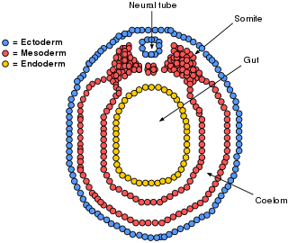
The mesoderm is the middle layer of the three germ layers that develops during gastrulation in the very early development of the embryo of most animals. The outer layer is the ectoderm, and the inner layer is the endoderm.

<span class="mw-page-title-main">Peritoneum</span> Serous membrane that forms lining of abdominal cavity or coelom

The peritoneum is the serous membrane forming the lining of the abdominal cavity or coelom in amniotes and some invertebrates, such as annelids. It covers most of the intra-abdominal organs, and is composed of a layer of mesothelium supported by a thin layer of connective tissue. This peritoneal lining of the cavity supports many of the abdominal organs and serves as a conduit for their blood vessels, lymphatic vessels, and nerves.

<span class="mw-page-title-main">Abdominal cavity</span> Body cavity in the abdominal area

The abdominal cavity is a large body cavity in humans and many other animals that contain organs. It is a part of the abdominopelvic cavity. It is located below the thoracic cavity, and above the pelvic cavity. Its dome-shaped roof is the thoracic diaphragm, a thin sheet of muscle under the lungs, and its floor is the pelvic inlet, opening into the pelvis.

<span class="mw-page-title-main">Body cavity</span> Internal space within a multicellular organism

A body cavity is any space or compartment, or potential space, in an animal body. Cavities accommodate organs and other structures; cavities as potential spaces contain fluid.

<span class="mw-page-title-main">Coelom</span> The main body cavity in many animals

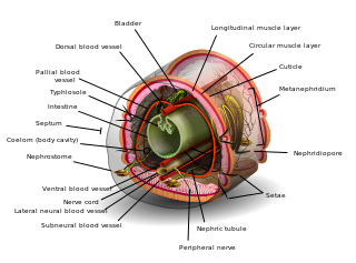
The coelom is the main body cavity in many animals and is positioned inside the body to surround and contain the digestive tract and other organs. In some animals, it is lined with mesothelium. In other animals, such as molluscs, it remains undifferentiated. In the past, and for practical purposes, coelom characteristics have been used to classify bilaterian animal phyla into informal groups.

<span class="mw-page-title-main">Pleural cavity</span> Thin fluid-filled space between the two pulmonary pleurae (visceral and parietal) of each lung

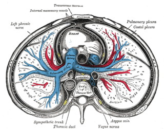
The pleural cavity, pleural space, or interpleural space is the potential space between the pleurae of the pleural sac that surrounds each lung. A small amount of serous pleural fluid is maintained in the pleural cavity to enable lubrication between the membranes, and also to create a pressure gradient.

<span class="mw-page-title-main">Pericardium</span> Double-walled sac containing the heart and roots of the great vessels

The pericardium, also called pericardial sac, is a double-walled sac containing the heart and the roots of the great vessels. It has two layers, an outer layer made of strong inelastic connective tissue, and an inner layer made of serous membrane. It encloses the pericardial cavity, which contains pericardial fluid, and defines the middle mediastinum. It separates the heart from interference of other structures, protects it against infection and blunt trauma, and lubricates the heart's movements.

<span class="mw-page-title-main">Respiratory tract</span> Organs involved in transmission of air to and from the point where gases diffuse into tissue

The respiratory tract is the subdivision of the respiratory system involved with the process of respiration in mammals. The respiratory tract is lined with respiratory epithelium as respiratory mucosa.

<span class="mw-page-title-main">Mesothelium</span> Membrane lining body cavities

The mesothelium is a membrane composed of simple squamous epithelial cells of mesodermal origin, which forms the lining of several body cavities: the pleura, peritoneum and pericardium.

The peritoneal cavity is a potential space between the parietal peritoneum and visceral peritoneum. The parietal and visceral peritonea are layers of the peritoneum named depending on their function/location. It is one of the spaces derived from the coelomic cavity of the embryo, the others being the pleural cavities around the lungs and the pericardial cavity around the heart.

A germ layer is a primary layer of cells that forms during embryonic development. The three germ layers in vertebrates are particularly pronounced; however, all eumetazoans produce two or three primary germ layers. Some animals, like cnidarians, produce two germ layers making them diploblastic. Other animals such as bilaterians produce a third layer between these two layers, making them triploblastic. Germ layers eventually give rise to all of an animal's tissues and organs through the process of organogenesis.

<span class="mw-page-title-main">Gestational sac</span> Cavity of fluid surrounding an embryo

The gestational sac is the large cavity of fluid surrounding the embryo. During early embryogenesis it consists of the extraembryonic coelom, also called the chorionic cavity. The gestational sac is normally contained within the uterus. It is the only available structure that can be used to determine if an intrauterine pregnancy exists until the embryo can be identified.

In physiology, serous fluid or serosal fluid is any of various body fluids resembling serum, that are typically pale yellow or transparent and of a benign nature. The fluid fills the inside of body cavities. Serous fluid originates from serous glands, with secretions enriched with proteins and water. Serous fluid may also originate from mixed glands, which contain both mucous and serous cells. A common trait of serous fluids is their role in assisting digestion, excretion, and respiration.

<span class="mw-page-title-main">Lateral plate mesoderm</span>

The lateral plate mesoderm is the mesoderm that is found at the periphery of the embryo. It is to the side of the paraxial mesoderm, and further to the axial mesoderm. The lateral plate mesoderm is separated from the paraxial mesoderm by a narrow region of intermediate mesoderm. The mesoderm is the middle layer of the three germ layers, between the outer ectoderm and inner endoderm.

<span class="mw-page-title-main">Bilaminar embryonic disc</span>

The bilaminar embryonic disc, bilaminar blastoderm or embryonic disc is the distinct two-layered structure of cells formed in an embryo. In the development of the human embryo this takes place by day eight. It is formed when the inner cell mass, also known as the embryoblast, forms a bilaminar disc of two layers, an upper layer called the epiblast and a lower layer called the hypoblast, which will eventually form into fetus. These two layers of cells are stretched between two fluid-filled cavities at either end: the primitive yolk sac and the amniotic sac.

<span class="mw-page-title-main">Human embryonic development</span> Development and formation of the human embryo

Human embryonic development or human embryogenesis is the development and formation of the human embryo. It is characterised by the processes of cell division and cellular differentiation of the embryo that occurs during the early stages of development. In biological terms, the development of the human body entails growth from a one-celled zygote to an adult human being. Fertilization occurs when the sperm cell successfully enters and fuses with an egg cell (ovum). The genetic material of the sperm and egg then combine to form the single cell zygote and the germinal stage of development commences. Embryonic development in the human, covers the first eight weeks of development; at the beginning of the ninth week the embryo is termed a fetus. The eight weeks has 23 stages.

In the development of the human embryo, the intraembryonic coelom is a portion of the conceptus forming in the mesoderm during the third week of development. During the third week of development, the lateral plate mesoderm splits into a dorsal somatic mesoderm (somatopleure) and a ventral splanchnic mesoderm (splanchnopleure). The resulting cavity between the somatopleure and splanchnopleure is called the intraembryonic coelom. This space will give rise to the thoracic and abdominal cavities. The coelomic spaces in the lateral mesoderm and cardiogenic area are isolated. The isolated coelom begins to organize into a horseshoe shape. The spaces soon join together and form a single horseshoe-shaped cavity: the intraembryonic coelom. It then separates the mesoderm into two layers.

The development of the digestive system in the human embryo concerns the epithelium of the digestive system and the parenchyma of its derivatives, which originate from the endoderm. Connective tissue, muscular components, and peritoneal components originate in the mesoderm. Different regions of the gut tube such as the esophagus, stomach, duodenum, etc. are specified by a retinoic acid gradient that causes transcription factors unique to each region to be expressed. Differentiation of the gut and its derivatives depends upon reciprocal interactions between the gut endoderm and its surrounding mesoderm. Hox genes in the mesoderm are induced by a Hedgehog signaling pathway secreted by gut endoderm and regulate the craniocaudal organization of the gut and its derivatives. The gut system extends from the oropharyngeal membrane to the cloacal membrane and is divided into the foregut, midgut, and hindgut.

<span class="mw-page-title-main">Pulmonary pleurae</span> Serous membrane that lines the wall of the thoracic cavity and the surface of the lung

The pulmonary pleurae are the two opposing layers of serous membrane overlying the lungs, mediastinum and the inside surfaces of the surrounding chest walls.

A tissue membrane is a thin layer or sheet of cells that covers the outside of the body, the organs, internal passageways that lead to the exterior of the body, and the lining of the moveable joint cavities. There are two basic types of tissue membranes: connective tissue and epithelial membranes.

References

This Wikipedia entry incorporates text from the freely licensed Connexions edition of Anatomy & Physiology text-book by OpenStax College

  1. J. Gordon Betts; Kelly A. Young; James A. Wise; Eddie Johnson; Brandon Poe; Dean H. Kruse; Oksana Korol; Jody E. Johnson; Mark Womble; Peter DeSaix (Apr 25, 2013). "1.6 Anatomical Terminology". Anatomy and Physiology. Houston, Texas: OpenStax.
  2. Tank PW (2013). "Chapter 4: The abdomen". Grant's dissector (Fifteenth ed.). Philadelphia. ISBN   978-1-60913-606-2.{{cite book}}: CS1 maint: location missing publisher (link)
  3. "The Anatomy of Lining and Covering Tissues-Membranes!". McGraw-Hill Companies. Retrieved September 2, 2017.
  4. 1 2 3 4 "Anatomy & Physiology". Openstax college at Connexions. Retrieved November 16, 2013.