Pterygomandibular raphe

Last updated
Pterygomandibular raphe
Gray380.png
Muscles of the pharynx and cheek. (Pterygo-Mandibular ligament labeled at center, vertically.)
Details
Part of Buccopharyngeal fascia
Origin Pterygoid hamulus of the medial pterygoid plate
Insertion Mylohyoid line of the mandible
Identifiers
Latin raphe pterygomandibularis
TA98 A05.3.01.102
TA2 2178
FMA 55618
Anatomical terminology

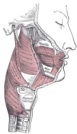
The pterygomandibular raphe (pterygomandibular fold [1] or pterygomandibular ligament) is a thin [2] tendinous band of buccopharyngeal fascia. It is attached superiorly to the pterygoid hamulus of the medial pterygoid plate, and inferiorly to the posterior end of the mylohyoid line of the mandible. It gives attachment to the buccinator muscle (in front), and the superior pharyngeal constrictor muscle (behind).

Contents

Structure

Open mouth, view from front. The pterygomandibular ligaments are marked with arrows. Note that in this mouth the lower wisdom teeth have been removed, slightly changing the shape of the pterygomandibular ligaments that usually curve and attach to the mandible around them. Open mouth pterygomandibular ligaments.png
Open mouth, view from front. The pterygomandibular ligaments are marked with arrows. Note that in this mouth the lower wisdom teeth have been removed, slightly changing the shape of the pterygomandibular ligaments that usually curve and attach to the mandible around them.

The pterygomandibular raphe is a tendinous band [2] [3] formed by the buccopharyngeal fascia. It is a paired structure, with one on each side of the mouth. [3] Superiorly, it is attached to the pterygoid hamulus of the medial pterygoid plate of the sphenoid bone; inferiorly, it is attached to the posterior end of the mylohyoid line of the mandible. [2] [3]

Relations

The pterygomandibular raphe is the common meeting point of the superior pharyngeal constrictor muscle and the buccinator muscle. This common attachment makes the raphe a junction of the oral cavity, oropharynx, and nasopharynx. [3]

The inferior alveolar nerve passes lateral to the raphe; the raphe is therefore a landmark for a nerve block of this nerve. [2]

The general location of the raphe is indicated by the pterygomandibular fold. [3]

Attachments

The pterygomandibular raphe gives attachment to the central portion of the buccinator muscle anteriorly, and to the superior pharyngeal constrictor muscle posteriorly. [2]

Variation

In foetuses, the pterygomandibular raphe is always very prominent. However, in adults, it may become less distinctive. It is very large and distinctive, in around 36% adults. It is fairly small, and only an upper triangular portion visible, in around 36% of adults. It is not visible in around 28%, making the superior pharyngeal constrictor muscle and the buccinator muscle continuous. This may vary by ethnic group. [4]

Clinical significance

When the mandible is splinted for gradual realignment (such as to treat sleep apnea), the pterygomandibular ligament slightly resists the realignment. [5]

The raphe is a landmark for administration of inferior alveolar nerve blocks. [2] [3] [1]

History

The pterygomandibular ligament was first noted in 1784. [3]

See also

Related Research Articles

Articles related to anatomy include:

<span class="mw-page-title-main">Mylohyoid muscle</span> Paired muscle of the neck

The mylohyoid muscle or diaphragma oris is a paired muscle of the neck. It runs from the mandible to the hyoid bone, forming the floor of the oral cavity of the mouth. It is named after its two attachments near the molar teeth. It forms the floor of the submental triangle. It elevates the hyoid bone and the tongue, important during swallowing and speaking.

<span class="mw-page-title-main">Superior pharyngeal constrictor muscle</span> Muscle

The superior pharyngeal constrictor muscle is a quadrilateral muscle of the pharynx. It is the uppermost and thinnest of the three pharyngeal constrictors.

<span class="mw-page-title-main">Stylopharyngeus muscle</span> Muscle

The stylopharyngeus muscle is a muscle in the head. It originates from the temporal styloid process. Some of its fibres insert onto the thyroid cartilage, while others end by intermingling with proximal structures. It is innervated by the glossopharyngeal nerve. It acts to elevate the larynx and pharynx, and dilate the pharynx, thus facilitating swallowing.

<span class="mw-page-title-main">Mylohyoid line</span> Bony ridge on the inner surface of the body of the mandible

The mylohyoid line is a bony ridge on the internal surface of the mandible. It runs posterosuperiorly. It is the site of origin of the mylohyoid muscle, the superior pharyngeal constrictor muscle, and the pterygomandibular raphe.

<span class="mw-page-title-main">Sphenomandibular ligament</span>

The sphenomandibular ligament is one of the three ligaments of the temporomandibular joint. It is situated medially to - and generally separate from - the articular capsule of the joint. Superiorly, it is attached to the spine of the sphenoid bone; inferiorly, it is attached to the lingula of mandible. The SML acts to limit inferior-ward movement of the mandible.

<span class="mw-page-title-main">Pterygoid processes of the sphenoid</span> Bone plates projecting from the sphenoid bone of the skull

The pterygoid processes of the sphenoid, one on either side, descend perpendicularly from the regions where the body and the greater wings of the sphenoid bone unite.

<span class="mw-page-title-main">Maxillary artery</span> Artery supplying face structures in humans

The maxillary artery supplies deep structures of the face. It branches from the external carotid artery just deep to the neck of the mandible.

<span class="mw-page-title-main">Infratemporal fossa</span> Cavity that is part of the skull

The infratemporal fossa is an irregularly shaped cavity that is a part of the skull. It is situated below and medial to the zygomatic arch. It is not fully enclosed by bone in all directions. It contains superficial muscles, including the lower part of the temporalis muscle, the lateral pterygoid muscle, and the medial pterygoid muscle. It also contains important blood vessels such as the middle meningeal artery, the pterygoid plexus, and the retromandibular vein, and nerves such as the mandibular nerve (CN V3) and its branches.

<span class="mw-page-title-main">Anterior triangle of the neck</span>

The anterior triangle is a region of the neck.

<span class="mw-page-title-main">Deep cervical fascia</span>

The deep cervical fascia lies under cover of the platysma, and invests the muscles of the neck; it also forms sheaths for the carotid vessels, and for the structures situated in front of the vertebral column. Its attachment to the hyoid bone prevents the formation of a dewlap.

<span class="mw-page-title-main">Prevertebral fascia</span> Layer of deep cervical fascia that surrounds the vertebral column

The prevertebral fascia is the layer of deep cervical fascia that surrounds the vertebral column. It is the deepest layer of deep cervical fascia.

<span class="mw-page-title-main">Buccopharyngeal fascia</span>

The buccopharyngeal fascia is a fascia of the pharynx. It represents the posterior portion of the pretracheal fascia. It covers the superior pharyngeal constrictor muscles, and buccinator muscle.

The pharyngobasilar fascia is a fascia of the pharynx. It is situated between the mucous and muscular layers of the pharynx. It is formed as a thickening of the pharyngeal mucosa superior to the superior pharyngeal constrictor muscle. It attaches to the basilar part of occipital bone, the petrous part of the temporal bone, the medial pterygoid plate, and the pterygomandibular raphe. It diminishes in thickness inferiorly. Posteriorly, it is reinforced by the pharyngeal raphe. It reinforces the pharyngeal wall where muscle is deficient.

<span class="mw-page-title-main">Outline of human anatomy</span> Overview of and topical guide to human anatomy

The following outline is provided as an overview of and topical guide to human anatomy:

<span class="mw-page-title-main">Parapharyngeal space</span>

The parapharyngeal space, is a potential space in the head and the neck. It has clinical importance in otolaryngology due to parapharyngeal space tumours and parapharyngeal abscess developing in this area. It is also a key anatomic landmark for localizing disease processes in the surrounding spaces of the neck; the direction of its displacement indirectly reflects the site of origin for masses or infection in adjacent areas, and consequently their appropriate differential diagnosis.

<span class="mw-page-title-main">Mandible</span> Lower jaw bone

In jawed vertebrates, the mandible, lower jaw, or jawbone is a bone that makes up the lower – and typically more mobile – component of the mouth.

<span class="mw-page-title-main">Submandibular space</span>

The submandibular space is a fascial space of the head and neck. It is a potential space, and is paired on either side, located on the superficial surface of the mylohyoid muscle between the anterior and posterior bellies of the digastric muscle. The space corresponds to the anatomic region termed the submandibular triangle, part of the anterior triangle of the neck.

Fascial spaces are potential spaces that exist between the fasciae and underlying organs and other tissues. In health, these spaces do not exist; they are only created by pathology, e.g. the spread of pus or cellulitis in an infection. The fascial spaces can also be opened during the dissection of a cadaver. The fascial spaces are different from the fasciae themselves, which are bands of connective tissue that surround structures, e.g. muscles. The opening of fascial spaces may be facilitated by pathogenic bacterial release of enzymes which cause tissue lysis. The spaces filled with loose areolar connective tissue may also be termed clefts. Other contents such as salivary glands, blood vessels, nerves and lymph nodes are dependent upon the location of the space. Those containing neurovascular tissue may also be termed compartments.

<span class="mw-page-title-main">Pterygomandibular space</span>

The pterygomandibular space is a fascial space of the head and neck. It is a potential space in the head and is paired on each side. It is located between the lateral pterygoid muscle and the medial surface of the ramus of the mandible. The pterygomandibular space is one of the four compartments of the masticator space.

References

  1. 1 2 Fehrenbach, Margaret J.; Herring, Susan W. (2017). Illustrated Anatomy of the Head and Neck (5th ed.). St. Louis: Elsevier. p. 267. ISBN   978-0-323-39634-9.
  2. 1 2 3 4 5 6 7 8 Standring, Susan (2020). Gray's Anatomy: The Anatomical Basis of Clinical Practice (42th ed.). New York. p. 625. ISBN   978-0-7020-7707-4. OCLC   1201341621.{{cite book}}: CS1 maint: location missing publisher (link)
  3. 1 2 3 4 5 6 7 8 9 10 Rao, D.; Sandhu, S.J.S.; Ormsby, C.; Natter, P.; Haymes, D.; Cohen, I.; Jenson, M. (2017-04-01). "Review of the Pterygomandibular Raphe". Neurographics. 7 (2): 121–125. doi:10.3174/ng.2170196.
  4. Shimada, Kazuyuki; Gasser, Raymond F. (1989). "Morphology of the pterygomandibular raphe in human fetuses and adults". The Anatomical Record. 224 (1): 117–122. doi:10.1002/ar.1092240115. ISSN   1097-0185. PMID   2729614. S2CID   36677456.
  5. Brown, Elizabeth C; Jugé, Lauriane; Knapman, Fiona L; Burke, Peter G R; Ngiam, Joachim; Sutherland, Kate; Butler, Jane E; Eckert, Danny J; Cistulli, Peter A; Bilston, Lynne E (2021-04-01). "Mandibular advancement splint response is associated with the pterygomandibular raphe". Sleep. 44 (4). doi: 10.1093/sleep/zsaa222 . ISSN   0161-8105. PMID   33146716.