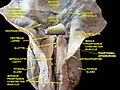
Pharyngobasilar fascia

Last updated
Pharyngobasilar fascia
Details
Identifiers
Latin fascia pharyngobasilaris
TA98 A05.3.01.027
TA2 2858
FMA 55074
Anatomical terminology

The pharyngobasilar fascia is a fascia of the pharynx. [1] It is situated between the mucous and muscular layers of the pharynx.[ citation needed ] It is formed as a thickening of the pharyngeal mucosa superior to the superior pharyngeal constrictor muscle. It attaches to the basilar part of occipital bone, the petrous part of the temporal bone (medial to the pharyngotympanic tube), the (posterior border of the) medial pterygoid plate, and the pterygomandibular raphe. It diminishes in thickness inferiorly. Posteriorly, it is reinforced by the pharyngeal raphe. [1] It reinforces the pharyngeal wall where muscle is deficient.[ citation needed ]

Contents

Additional images

See also

Related Research Articles

<span class="mw-page-title-main">Tibia</span> Leg bone in vertebrates

The tibia, also known as the shinbone or shankbone, is the larger, stronger, and anterior (frontal) of the two bones in the leg below the knee in vertebrates ; it connects the knee with the ankle. The tibia is found on the medial side of the leg next to the fibula and closer to the median plane. The tibia is connected to the fibula by the interosseous membrane of leg, forming a type of fibrous joint called a syndesmosis with very little movement. The tibia is named for the flute tibia. It is the second largest bone in the human body, after the femur. The leg bones are the strongest long bones as they support the rest of the body.

<span class="mw-page-title-main">Hyoid bone</span> Bone situated in the neck between the chin and the thyroid cartilage

The hyoid bone is a horseshoe-shaped bone situated in the anterior midline of the neck between the chin and the thyroid cartilage. At rest, it lies between the base of the mandible and the third cervical vertebra.

<span class="mw-page-title-main">External carotid artery</span> Major artery of the head and neck

The external carotid artery is a major artery of the head and neck. It arises from the common carotid artery when it splits into the external and internal carotid artery. The external carotid artery supplies blood to the face, brain and neck.

<span class="mw-page-title-main">Mylohyoid muscle</span> Paired muscle of the neck

The mylohyoid muscle or diaphragma oris is a paired muscle of the neck. It runs from the mandible to the hyoid bone, forming the floor of the oral cavity of the mouth. It is named after its two attachments near the molar teeth. It forms the floor of the submental triangle. It elevates the hyoid bone and the tongue, important during swallowing and speaking.

<span class="mw-page-title-main">Common carotid artery</span> One of the two arteries that supply the head and neck with blood

In anatomy, the left and right common carotid arteries (carotids) are arteries that supply the head and neck with oxygenated blood; they divide in the neck to form the external and internal carotid arteries.

<span class="mw-page-title-main">Sternothyroid muscle</span>

The sternothyroid muscle is an infrahyoid muscle of the neck. It acts to depress the hyoid bone.

<span class="mw-page-title-main">Levator veli palatini</span> Muscle of the soft palate

The levator veli palatini is a muscle of the soft palate and pharynx. It is innervated by the vagus nerve via its pharyngeal plexus. During swallowing, it contracts, elevating the soft palate to help prevent food from entering the nasopharynx.

<span class="mw-page-title-main">Salpingopharyngeus muscle</span> Muscle of the pharynx

The salpingopharyngeus muscle is a muscle of the pharynx. It arises from the lower part of the cartilage of the Eustachian tube, and inserts into the palatopharyngeus muscle by blending with its posterior fasciculus. It is innervated by vagus nerve via the pharyngeal plexus. It raises the pharynx and larynx during deglutition (swallowing) and laterally draws the pharyngeal walls up. It opens the pharyngeal orifice of the Eustachian tube during swallowing to allow for the equalization of pressure between it and the pharynx.

<span class="mw-page-title-main">Middle pharyngeal constrictor muscle</span> Muscle in the neck

The middle pharyngeal constrictor is a fan-shaped muscle located in the neck. It is one of three pharyngeal constrictor muscles. It is smaller than the inferior pharyngeal constrictor muscle.

<span class="mw-page-title-main">Inferior pharyngeal constrictor muscle</span> Skeletal muscle of the pharynx

The inferior pharyngeal constrictor muscle is a skeletal muscle of the neck. It is the thickest of the three outer pharyngeal muscles. It arises from the sides of the cricoid cartilage and the thyroid cartilage. It is supplied by the vagus nerve. It is active during swallowing, and partially during breathing and speech. It may be affected by Zenker's diverticulum.

<span class="mw-page-title-main">Superior pharyngeal constrictor muscle</span> Muscle

The superior pharyngeal constrictor muscle is a quadrilateral muscle of the pharynx. It is the uppermost and thinnest of the three pharyngeal constrictors.

<span class="mw-page-title-main">Stylopharyngeus muscle</span> Muscle

The stylopharyngeus muscle is a muscle in the head. It originates from the temporal styloid process. Some of its fibres insert onto the thyroid cartilage, while others end by intermingling with proximal structures. It is innervated by the glossopharyngeal nerve. It acts to elevate the larynx and pharynx, and dilate the pharynx, thus facilitating swallowing.

<span class="mw-page-title-main">Superior thyroid artery</span>

The superior thyroid artery arises from the external carotid artery just below the level of the greater cornu of the hyoid bone and ends in the thyroid gland.

<span class="mw-page-title-main">Inferior thyroid artery</span> Artery of the neck

The inferior thyroid artery is an artery in the neck. It arises from the thyrocervical trunk and passes upward, in front of the vertebral artery and longus colli muscle. It then turns medially behind the carotid sheath and its contents, and also behind the sympathetic trunk, the middle cervical ganglion resting upon the vessel.

<span class="mw-page-title-main">Carotid triangle</span>

The carotid triangle is a portion of the anterior triangle of the neck.

<span class="mw-page-title-main">Pterygomandibular raphe</span> Ligament formed from the buccopharyngeal fascia

The pterygomandibular raphe is a thin tendinous band of buccopharyngeal fascia. It is attached superiorly to the pterygoid hamulus of the medial pterygoid plate, and inferiorly to the posterior end of the mylohyoid line of the mandible. It gives attachment to the buccinator muscle, and the superior pharyngeal constrictor muscle (behind).

<span class="mw-page-title-main">Prevertebral fascia</span> Layer of deep cervical fascia that surrounds the vertebral column

The prevertebral fascia is the layer of deep cervical fascia that surrounds the vertebral column. It is the deepest layer of deep cervical fascia.

<span class="mw-page-title-main">Buccopharyngeal fascia</span>

The buccopharyngeal fascia is a fascia of the pharynx. It represents the posterior portion of the pretracheal fascia. It covers the superior pharyngeal constrictor muscles, and buccinator muscle.

<span class="mw-page-title-main">Pharynx</span> Part of the throat that is behind the mouth and nasal cavity

The pharynx is the part of the throat behind the mouth and nasal cavity, and above the esophagus and trachea. It is found in vertebrates and invertebrates, though its structure varies across species. The pharynx carries food to the esophagus and air to the larynx. The flap of cartilage called the epiglottis stops food from entering the larynx.

<span class="mw-page-title-main">Parapharyngeal space</span>

The parapharyngeal space, is a potential space in the head and the neck. It has clinical importance in otolaryngology due to parapharyngeal space tumours and parapharyngeal abscess developing in this area. It is also a key anatomic landmark for localizing disease processes in the surrounding spaces of the neck; the direction of its displacement indirectly reflects the site of origin for masses or infection in adjacent areas, and consequently their appropriate differential diagnosis.

References

  1. 1 2 Standring, Susan (2020). Gray's Anatomy: The Anatomical Basis of Clinical Practice (42nd ed.). New York. p. 709. ISBN   978-0-7020-7707-4. OCLC   1201341621.{{cite book}}: CS1 maint: location missing publisher (link)