Deep cervical fascia

Last updated
Deep cervical fascia
Gray384.png
Section of the neck at about the level of the sixth cervical vertebra. Showing the arrangement of the fascia colli.
Anatomical terminology

The deep cervical fascia (or fascia colli in older texts) lies under cover of the platysma, and invests the muscles of the neck; it also forms sheaths for the carotid vessels, and for the structures situated in front of the vertebral column. Its attachment to the hyoid bone prevents the formation of a dewlap. [1]

Contents

The investing portion of the fascia is attached behind to the ligamentum nuchæ and to the spinous process of the seventh cervical vertebra.

The alar fascia is a portion of the deep cervical fascia.

Divisions

The deep cervical fascia is often divided into a superficial, middle, and deep layer.

The superficial layer is also known as the investing layer of deep cervical fascia. It envelops the trapezius, sternocleidomastoid, and muscles of facial expression. It also contains the submandibular and parotid salivary gland as well as the muscles of mastication (the masseter, pterygoid, and temporalis muscles).

The middle layer is also known as the pretracheal fascia. It envelopes the strap muscles (sternohyoid, sternothyroid, thyrohyoid, and omohyoid muscles). It also surrounds the pharynx, larynx, trachea, esophagus, thyroid, parathyroids, buccinators, and constrictor muscles of the pharynx.

The deep layer is also known as the prevertebral fascia. It surrounds the paraspinous muscles and cervical vertebrae. [2]

The carotid sheath is also considered a component of the deep cervical fascia.

Superior extent of the investing fascia

Above, the fascia is attached to the superior nuchal line of the occipital bone, to the mastoid process of the temporal bone, and to the whole length of the inferior border of the body of the mandible. [3]

Opposite the angle of the mandible the fascia is very strong, and binds the anterior edge of the sternocleidomastoideus firmly to that bone.

Between the mandible and the mastoid process it ensheathes the parotid gland—the layer which covers the gland extends upward under the name of the parotideomasseteric fascia and is fixed to the zygomatic arch. It also contributes to the sheath of the digastric.

At the level of the jaw, it splits to enclose the submandibular gland, with the upper leaflet inserting on the mylohyoid line just inferior to mylohyoid and the inferior leaflet inserting onto the lower margin of the jaw. The posterior portion of the upper leaflet helps separate the parotid gland from the submandibular gland where the mylohyoid is deficient while the posterior border is thickened into a strong band extending between the angle of the jaw and the temporal styloid process, forming the stylomandibular ligament. It is complemented by the pterygospinous ligament, which stretches from the upper part of the posterior border of the lateral pterygoid plate to the spinous process of the sphenoid. It occasionally ossifies, and in such cases, between its upper border and the base of the skull, a foramen is formed which transmits the branches of the mandibular nerve to the muscles of mastication.

Inferior extent of the investing fascia

Below, the fascia is attached to the thoracic outlet (acromion, clavicle, and manubrium). In doing so, it bifurcates into two layers, superficial and deep.

The former is attached to the anterior border of the manubrium, the latter to its posterior border and to the interclavicular ligament.

Between these two layers is a slit-like interval, the suprasternal space (space of Burns); it contains a small quantity of areolar tissue, the lower portions of the anterior jugular veins and their transverse connecting branch, the sternal heads of the sternocleidomastoid, and sometimes a lymph gland.

Deeper fascial layers

The fascia which lines the deep surface of the sternocleidomastoideus gives off the following processes:

Related Research Articles

<span class="mw-page-title-main">Neck</span> Body part that connects the head and torso

The neck is the part of the body on many vertebrates that connects the head with the torso. The neck supports the weight of the head and protects the nerves that carry sensory and motor information from the brain down to the rest of the body. In addition, the neck is highly flexible and allows the head to turn and flex in all directions. The structures of the human neck are anatomically grouped into four compartments: vertebral, visceral and two vascular compartments. Within these compartments, the neck houses the cervical vertebrae and cervical part of the spinal cord, upper parts of the respiratory and digestive tracts, endocrine glands, nerves, arteries and veins. Muscles of the neck are described separately from the compartments. They bound the neck triangles.

Articles related to anatomy include:

<span class="mw-page-title-main">Great auricular nerve</span> Cutaneous nerve of the head

The great auricular nerve is a cutaneous (sensory) nerve of the head. It originates from the second and third cervical (spinal) nerves (C2-C3) of the cervical plexus. It provides sensory innervation to the skin over the parotid gland and the mastoid process, parts of the outer ear, and to the parotid gland and its fascia.

<span class="mw-page-title-main">Parotid gland</span> Major salivary gland in many animals

The parotid gland is a major salivary gland in many animals. In humans, the two parotid glands are present on either side of the mouth and in front of both ears. They are the largest of the salivary glands. Each parotid is wrapped around the mandibular ramus, and secretes serous saliva through the parotid duct into the mouth, to facilitate mastication and swallowing and to begin the digestion of starches. There are also two other types of salivary glands; they are submandibular and sublingual glands. Sometimes accessory parotid glands are found close to the main parotid glands.

<span class="mw-page-title-main">Submandibular gland</span> Human salivary gland

The paired submandibular glands are major salivary glands located beneath the floor of the mouth. In adult humans, they each weigh about 15 grams and contribute some 60–67% of unstimulated saliva secretion; on stimulation their contribution decreases in proportion as parotid gland secretion rises to 50%. The average length of the normal adult human submandibular salivary gland is approximately 27 mm, while the average width is approximately 14.3 mm.

<span class="mw-page-title-main">Digastric muscle</span> Small muscle located under the jaw in mammals

The digastric muscle is a bilaterally paired suprahyoid muscle located under the jaw. Its posterior belly is attached to the mastoid notch of temporal bone, and its anterior belly is attached to the digastric fossa of mandible; the two bellies are united by an intermediate tendon which is held in a loop that attaches to the hyoid bone. The anterior belly is innervated via the mandibular nerve, and the posterior belly is innervated via the facial nerve. It may act to depress the mandible or elevate the hyoid bone.

<span class="mw-page-title-main">Common carotid artery</span> One of the two arteries that supply the head and neck with blood

In anatomy, the left and right common carotid arteries (carotids) are arteries that supply the head and neck with oxygenated blood; they divide in the neck to form the external and internal carotid arteries.
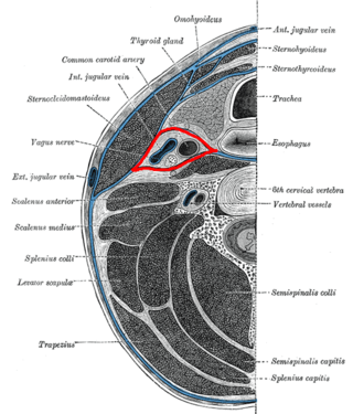
<span class="mw-page-title-main">Carotid sheath</span> Part of neck anatomy

The carotid sheath is a condensation of the deep cervical fascia enveloping multiple vital neurovascular structures of the neck, including the common and internal carotid arteries, the internal jugular vein, the vagus nerve, and ansa cervicalis. The carotid sheath helps protects the structures contained therein.

<span class="mw-page-title-main">Occipital artery</span>

The occipital artery is a branch of the external carotid artery that provides arterial supply to the back of the scalp, sternocleidomastoid muscles, and deep muscles of the back and neck.

<span class="mw-page-title-main">Posterior auricular artery</span> Small artery

The posterior auricular artery is a small artery that arises from the external carotid artery. It ascends along the side of the head. It supplies several muscles of the neck and several structures of the head.

<span class="mw-page-title-main">Submandibular triangle</span>

The submandibular triangle corresponds to the region of the neck immediately beneath the body of the mandible.

<span class="mw-page-title-main">Carotid triangle</span>

The carotid triangle is a portion of the anterior triangle of the neck.
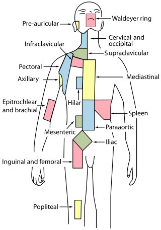
<span class="mw-page-title-main">Cervical lymph nodes</span> Lymph nodes found in the neck

Cervical lymph nodes are lymph nodes found in the neck. Of the 800 lymph nodes in the human body, 300 are in the neck. Cervical lymph nodes are subject to a number of different pathological conditions including tumours, infection and inflammation.
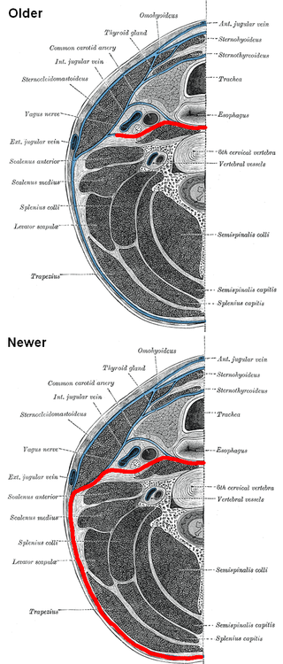
<span class="mw-page-title-main">Prevertebral fascia</span> Layer of deep cervical fascia that surrounds the vertebral column

The prevertebral fascia is the layer of deep cervical fascia that surrounds the vertebral column. It is the deepest layer of deep cervical fascia.

<span class="mw-page-title-main">Investing layer of deep cervical fascia</span>

The investing layer of deep cervical fascia is the most superficial part of the deep cervical fascia, and encloses the whole neck.

<span class="mw-page-title-main">Pretracheal fascia</span>

The pretracheal fascia is a layer of the deep cervical fascia at the front of the neck. It attaches to the hyoid bone above, and - extending down into the thorax - blends with the fibrous pericardium below. It encloses the thyroid gland and parathyroid glands, trachea, and esophagus. It extends medially in front of the carotid vessels. It assists in forming the carotid sheath.

<span class="mw-page-title-main">Outline of human anatomy</span> Overview of and topical guide to human anatomy

The following outline is provided as an overview of and topical guide to human anatomy:

<span class="mw-page-title-main">Parapharyngeal space</span>

The parapharyngeal space, is a potential space in the head and the neck. It has clinical importance in otolaryngology due to parapharyngeal space tumours and parapharyngeal abscess developing in this area. It is also a key anatomic landmark for localizing disease processes in the surrounding spaces of the neck; the direction of its displacement indirectly reflects the site of origin for masses or infection in adjacent areas, and consequently their appropriate differential diagnosis.

<span class="mw-page-title-main">Submandibular space</span>

The submandibular space is a fascial space of the head and neck. It is a potential space, and is paired on either side, located on the superficial surface of the mylohyoid muscle between the anterior and posterior bellies of the digastric muscle. The space corresponds to the anatomic region termed the submandibular triangle, part of the anterior triangle of the neck.

The parotid fascia is a tough fascia enclosing the parotid gland. It has a superficial layer and a deep layer.
References

PD-icon.svgThis article incorporates text in the public domain from page 388 of the 20th edition of Gray's Anatomy (1918)

  1. Anatomy & Physiology, 8th Edition, McGraw-Hill Co., 2008.
  2. Lee, K.J. (2012). Essential Otolaryngology (10 ed.). McGraw Hill. pp. 559–60. ISBN   978-0-07-176-147-5.
  3. Meyers, E. S. (Errol Solomon); MacPherson, R. K. (Ronald Kenneth) (1939-01-01). The arrangement of the deep cervical fascia / by E.S. Meyers and R.K. MacPherson. Papers (University of Queensland. Dept. of Anatomy); v. 1, no. 1. Sydney: Australasian Medical Publishing.