Platysma muscle

Last updated
Platysma muscle
Sobo 1909 267.png
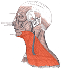
The platysma is visible, with skin removed.
Sobo 1909 260.png
The muscles of the face, platysma visible at bottom right.
Details
Origin subcutaneous tissue of infraclavicular and supraclavicular regions
Insertion base of mandible; skin of cheek and lower lip; angle of mouth; orbicularis oris
Artery branches of the submental artery and suprascapular artery
Nerve cervical branch of the facial nerve
Actions draws the corners of the mouth inferiorly and widens it (as in expressions of sadness and fright). Also draws the skin of the neck superiorly when teeth are clenched
Antagonist masseter muscle, temporalis muscle
Identifiers
Latin platysma
TA98 A04.2.01.001
TA2 2147
FMA 45738
Anatomical terms of muscle

The platysma muscle is a superficial muscle of the human neck that overlaps the sternocleidomastoid. It covers the anterior surface of the neck superficially. When it contracts, it produces a slight wrinkling of the neck, and a "bowstring" effect on either side of the neck.

Contents

Structure

The platysma muscle is a broad sheet of muscle arising from the fascia covering the upper parts of the pectoralis major muscle and deltoid muscle. Its fibers cross the clavicle, and proceed obliquely upward and medially along the side of the neck. This leaves the inferior part of the neck in the midline deficient of significant muscle cover. [1]

Fibres at the front of the muscle from the left and right sides intermingle together below and behind the mandibular symphysis, the junction where the two lateral halves of the mandible are fused at an early period of life (although not a true symphysis). Fibres at the back of the muscle cross the mandible, some being inserted into the bone below the oblique line, others into the skin and subcutaneous tissue of the lower part of the face. Many of these fibers blend with the muscles about the angle and lower part of the mouth. [1]

Sometimes fibers can be traced to the zygomaticus major muscle, or to the margin of the orbicularis oris muscle. Beneath the platysma, the external jugular vein descends from the angle of the mandible to the clavicle. [2]

Nerve supply

The platysma muscle is supplied by the cervical branch of the facial nerve. [1]

Blood supply

The platysma muscle is supplied by branches of the submental artery and suprascapular artery.

Relations

The platysma muscle lies just deep to the subcutaneous fascia and fat. [1] [3] It covers many structures found deeper in the neck, such as the external carotid artery, the external jugular vein, [4] the parotid gland, [4] the lesser occipital nerve, [4] the great auricular nerve, [4] and the marginal mandibular branch of the facial nerve. [5]

Variation

Variations occur in the extension over the face and over the clavicle and shoulder. The platysma muscle may be absent or interdigitate with the muscle of the opposite side in front of the neck; attachment to clavicle, mastoid process or occipital bone occurs. A more or less independent fasciculus, the occipitalis minor muscle, may extend from the fascia over the trapezius muscle to fascia over the insertion of the sternocleidomastoid muscle.

Function

Wrinkling

When the entire platysma muscle is in action, it produces a slight wrinkling of the surface of the skin of the neck in an oblique direction (at an angle to the midline). It creates a distinctive "bowstring" effect on either side of the neck, where fibres move away from the midline. [3]

Jaw and lip movement

The anterior portion of the platysma muscle, the thickest part of the muscle, depresses the lower jaw. It also draws down the lower lip and angle of the mouth in a frown. However, the platysma muscle plays only a minor role in depressing the lower lip, which is primarily performed by the depressor anguli oris muscle and the depressor labii inferioris muscle.

Clinical significance

In a similar fashion to other muscles, the platysma muscle is vulnerable to tears, strains and muscle atrophy, among many other possible conditions.

Injury

The platysma muscle is vulnerable to neck injuries that may penetrate it, as it is both superficial and thin. [6] Penetrating trauma in the neck injuries can be defined as any that completely penetrate the platysma muscle, making it an important landmark. [6] CTA (computed tomography angiography) may be used to visualise arteries and veins, such as for complex injuries from gunshot wounds or stab wounds, and is useful to image any damage to the muscle. [6] [7] This minimises the number of exploratory surgeries that need to be performed, thus improving the handling of the condition. [7]

Neck surgery

When neck surgery is performed, the platysma muscle usually needs to be cut through to access deeper structures. [1] [5] Fibres need to be sutured together accurately to prevent abnormal scar retraction, which may look unsightly. [1]

Plastic surgery

Wrinkly skin of neck caused by decrease in muscle tone leading to thinning and shortening of muscle is the secondary complication of facial nerve palsy and can be associated with the normal aging process. Neck bands in the area above the platysma muscle become most noticeable with age. [3] These may be aggravated by weightlifting or facelift procedures. This may be known as platysma dyskinesia or "turkey neck".

Conservative management may be used. Alternatively, interventions include botulinum toxin injection and platysmaplasty. Platysmaplasty is a surgery in this area, that can be open or closed; in the latter a specialised instrument called a plastymotome is used that allows the surgery to be done without incisions. [8] It takes approximately 2 weeks for the symptoms to be reduced. [9]

Adipose tissue is found above the platysma muscle, so liposuction of the neck may be performed fairly easily without the need to pierce it. [10] It is also important to not damage the platysma muscle to prevent bleeding. [10]

Images

Related Research Articles

<span class="mw-page-title-main">Subclavian artery</span> Major arteries of the upper thorax, below the clavicle

In human anatomy, the subclavian arteries are paired major arteries of the upper thorax, below the clavicle. They receive blood from the aortic arch. The left subclavian artery supplies blood to the left arm and the right subclavian artery supplies blood to the right arm, with some branches supplying the head and thorax. On the left side of the body, the subclavian comes directly off the aortic arch, while on the right side it arises from the relatively short brachiocephalic artery when it bifurcates into the subclavian and the right common carotid artery.

<span class="mw-page-title-main">Sternocleidomastoid muscle</span> Cervical muscle

The sternocleidomastoid muscle is one of the largest and most superficial cervical muscles. The primary actions of the muscle are rotation of the head to the opposite side and flexion of the neck. The sternocleidomastoid is innervated by the accessory nerve.

<span class="mw-page-title-main">Omohyoid muscle</span> Human neck muscle

The omohyoid muscle is a muscle in the neck. It is one of the infrahyoid muscles. It consists of two bellies separated by an intermediate tendon. Its inferior belly is attached to the scapula; its superior belly is attached to the hyoid bone. Its intermediate tendon is anchored to the clavicle and first rib by a fascial sling. The omohyoid is innervated by the ansa cervicalis of the cervical plexus. It acts to depress the hyoid bone.

<span class="mw-page-title-main">Stellate ganglion</span>

The stellate ganglion is a sympathetic ganglion formed by the fusion of the inferior cervical ganglion and the first thoracic ganglion, which is present in 80% of individuals. Sometimes, the second and the third thoracic ganglia are included in this fusion.

<span class="mw-page-title-main">Coccygeus muscle</span> Muscle of the lower back arising by its apex from the spine of the ischium

The coccygeus muscle or ischiococcygeus is a muscle of the pelvic floor, located posterior to levator ani and anterior to the sacrospinous ligament.

<span class="mw-page-title-main">Cricothyroid ligament</span>

The cricothyroid ligament is a ligament in the neck. It connects the cricoid cartilage to the thyroid cartilage. It prevents these cartilages from moving too far apart. It is cut during an emergency cricothyrotomy to treat upper airway obstruction.

<span class="mw-page-title-main">Mentalis</span> Muscle that raises the central portion of the lower lip

The mentalis muscle is a paired central muscle of the lower lip, situated at the tip of the chin. It originates from the mentum of the mandible, and inserts into the soft tissue of the chin. It is sometimes referred to as the "pouting muscle" due to it raising the lower lip and causing chin wrinkles.

<span class="mw-page-title-main">Facial artery</span>

The facial artery is a branch of the external carotid artery that supplies structures of the superficial face.

<span class="mw-page-title-main">Sternohyoid muscle</span>

The sternohyoid muscle is a bilaterally paired, long, thin, narrow strap muscle of the anterior neck. It is one of the infrahyoid muscles. It is innervated by the ansa cervicalis. It acts to depress the hyoid bone.

<span class="mw-page-title-main">Thyrohyoid muscle</span> Neck muscle that depresses the hyoid bone and elevates the larynx

The thyrohyoid muscle is a small skeletal muscle of the neck. Above, it attaches onto the greater cornu of the hyoid bone; below, it attaches onto the oblique line of the thyroid cartilage. It is innervated by fibres derived from the cervical spinal nerve 1 that run with the hypoglossal nerve to reach this muscle. The thyrohyoid muscle depresses the hyoid bone and elevates the larynx during swallowing. By controlling the position and shape of the larynx, it aids in making sound.

<span class="mw-page-title-main">Carotid sheath</span>

The carotid sheath is a condensation of the deep cervical fascia enveloping multiple vital neurovascular structures of the neck, including the common and internal carotid arteries, the internal jugular vein, the vagus nerve, and ansa cervicalis. The carotid sheath helps protects the structures contained therein.

<span class="mw-page-title-main">Occipitofrontalis muscle</span> Facial muscle helping to create facial expressions

The occipitofrontalis muscle is a muscle which covers parts of the skull. It consists of two parts or bellies: the occipital belly, near the occipital bone, and the frontal belly, near the frontal bone. It is supplied by the supraorbital artery, the supratrochlear artery, and the occipital artery. It is innervated by the facial nerve. In humans, the occipitofrontalis helps to create facial expressions.

<span class="mw-page-title-main">Lateral cutaneous nerve of thigh</span> Nerve of the thigh

The lateral cutaneous nerve of the thigh is a cutaneous nerve of the thigh. It originates from the dorsal divisions of the second and third lumbar nerves from the lumbar plexus. It passes under the inguinal ligament to reach the thigh. It supplies sensation to the skin on the lateral part of the thigh by an anterior branch and a posterior branch.

<span class="mw-page-title-main">Supraspinous ligament</span>

The supraspinous ligament, also known as the supraspinal ligament, is a ligament found along the vertebral column.

<span class="mw-page-title-main">Lingual artery</span>

The lingual artery arises from the external carotid artery between the superior thyroid artery and facial artery. It can be located easily in the tongue.

<span class="mw-page-title-main">Anterior jugular vein</span>

The anterior jugular vein is a vein in the neck.

<span class="mw-page-title-main">Lateral pectoral nerve</span>

The lateral pectoral nerve arises from the lateral cord of the brachial plexus, and through it from the C5-7.

<span class="mw-page-title-main">Retromandibular vein</span> Major face vein

The retromandibular vein is a major vein of the face. It is formed within the parotid gland by the confluence of the maxillary vein, and superficial temporal vein. It descends in the gland and splits into two branches upon emerging from the gland. Its anterior branch then joins the (anterior) facial vein forming the common facial vein, while its posterior branch joins the posterior auricular vein forming the external jugular vein.

<span class="mw-page-title-main">Infratemporal fossa</span> Cavity that is part of the skull

The infratemporal fossa is an irregularly shaped cavity that is a part of the skull. It is situated below and medial to the zygomatic arch. It is not fully enclosed by bone in all directions. It contains superficial muscles, including the lower part of the temporalis muscle, the lateral pterygoid muscle, and the medial pterygoid muscle. It also contains important blood vessels such as the middle meningeal artery, the pterygoid plexus, and the retromandibular vein, and nerves such as the mandibular nerve (CN V3) and its branches.

<span class="mw-page-title-main">Zygomatic branches of the facial nerve</span> Nerves of the face

The zygomatic branches of the facial nerve (malar branches) are nerves of the face. They run across the zygomatic bone to the lateral angle of the orbit. Here, they supply the orbicularis oculi muscle, and join with filaments from the lacrimal nerve and the zygomaticofacial branch of the maxillary nerve (CN V2).

References

PD-icon.svgThis article incorporates text in the public domain from page 387 of the 20th edition of Gray's Anatomy (1918)

  1. 1 2 3 4 5 6 Eibling, David E. (2008). "78 - Neck Dissection". Operative Otolaryngology: Head and Neck Surgery. Vol. 1 (2nd ed.). Philadelphia: Saunders. pp. 679–708. doi:10.1016/B978-1-4160-2445-3.50082-0. ISBN   978-1-4377-2083-9. OCLC   825780332.
  2. Kim, Se-Hoon; Chang, Ung-Kyu; Kim, Daniel H.; Bilsky, Mark H. (2006-01-01), Kim, Daniel H.; Chang, Ung-Kyu; Kim, Se-Hoon; Bilsky, Mark H. (eds.), "Chapter 19 - Management of Upper Cervical Spine Tumors", Tumors of the Spine, Philadelphia: W.B. Saunders, pp. 378–394, ISBN   978-1-4160-3367-7 , retrieved 2021-01-06
  3. 1 2 3 Posnick, Jeffrey C. (2014). "40 - Aesthetic Alteration of the Soft Tissues of the Neck and Lower Face: Evaluation and Surgery". Orthognathic Surgery: Principles & Practice. St. Louis: Saunders. pp. 1746–1783. doi:10.1016/B978-1-4557-2698-1.00040-X. ISBN   978-1-4557-5027-6. OCLC   860861780.
  4. 1 2 3 4 Kim, Se-Hoon; Chang, Ung-Kyu; Kim, Daniel H.; Bilsky, Mark H. (2008). "19 - Management of Upper Cervical Spine Tumors". Tumors of the Spine. Philadelphia: Saunders. pp. 378–394. doi:10.1016/B978-1-4160-3367-7.10019-7. ISBN   978-1-4377-2164-5. OCLC   489078564.
  5. 1 2 Wolfe, Michael J.; Wilson, Keith (2007). "21 - Head and Neck Cancer". Essentials of Surgical Oncology: Surgical Foundations. Philadelphia: Mosby. pp. 329–357. doi:10.1016/B978-0-8151-4385-7.50027-8. ISBN   978-0-8151-4385-7. OCLC   608607674.
  6. 1 2 3 Cothren, C. Clay; Moore, Ernest E. (2009). "19 - Penetrating Neck Trauma". Abernathy's Surgical Secrets (6th ed.). Philadelphia: Mosby. pp. 110–113. doi:10.1016/B978-0-323-05711-0.00019-7. ISBN   978-0-323-07475-9. OCLC   460933202.
  7. 1 2 Bell, RB; Osborn, T; Dierks, EJ; Potter, BE; Long, WB (2007). "Management of penetrating neck injuries: a new paradigm for civilian trauma". J. Oral Maxillofac. Surg. 65 (4): 691–705. doi:10.1016/j.joms.2006.04.044. PMID   17368366.
  8. Labb??n a similar fashion to other muscles, the platysma is vulnerable to tears, strains and muscle atrophy among many other possible conditions. The platysma is vulnerable to neck injuries that may penetrate it. A type of medical imaging called CTA (computed tomography angiography), used to visualise arterial and venous vessels, is useful to minimise the number of neck explorations, thus improving the handling of the condition.[3] Another area of importance of the platysma lies in plastic surgery. Neck bands in the area become most noticeable with age, aggravated by weightlifting or facelift. If it doesn't heal with time, there are many options to correct this: Botox/Dysport/Xeomin and platysmaplasty. Platysmaplasty is a surgery in this area, that can be open or closed, in the latter a specialised instrument called plastymotome that allow the surgery to be done without incisions. It takes approximately 2 weeks for the symptoms to be reduced.[4], Daniel; Franco, R G.; Nicolas, J (May 2006). "Platysma Suspension and Platysmaplasty during Neck Lift: Anatomical Study and Analysis of 30 Cases". Plastic and Reconstructive Surgery. 117 (6): 2001–2007. doi:10.1097/01.prs.0000218972.75144.9c. ISSN   0032-1052. PMID   16651976. S2CID   26104853.{{cite journal}}: CS1 maint: multiple names: authors list (link) CS1 maint: numeric names: authors list (link)
  9. Daher, JC (2011). "Closed platysmotomy: a new procedure for the treatment of platysma bands without skin dissection". Aesthetic Plast Surg. 35 (5): 866–77. doi:10.1007/s00266-011-9782-0. PMC   3192284 . PMID   21847680.
  10. 1 2 Alam, Murad; White, Lucile E. (2008). "1 - Anatomy in Dermatologic Surgery". Complications in Dermatologic Surgery. Philadelphia: Mosby. pp. 1–18. doi:10.1016/B978-0-323-04546-9.10001-9. ISBN   978-0-323-04546-9. OCLC   811786617.
Books