Depressor anguli oris muscle

Last updated
Depressor anguli oris
Gray381.png
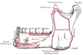
Scheme showing arrangement of fibers of Orbicularis oris (triangularis labeled at bottom right).
Depressor anguli oris.png
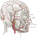
Muscles of the head, face, and neck (labeled as triangularis near chin).
Details
Origin tubercle of mandible
Insertion modiolus of mouth
Artery facial artery
Nerve marginal mandibular branch of the facial nerve
Actions depresses angle of mouth
Identifiers
Latin musculus depressor anguli oris
TA98 A04.1.03.026
TA2 2076
FMA 46828
Anatomical terms of muscle

The depressor anguli oris muscle (triangularis muscle) is a facial muscle. It originates from the mandible and inserts into the angle of the mouth. It is associated with frowning, as it depresses the corner of the mouth.

Contents

Structure

The depressor anguli oris arises from the lateral surface of the mandible. [1] Its fibers then converge. It is inserted by a narrow fasciculus into the angle of the mouth. [1] At its origin, it is continuous with the platysma muscle, and at its insertion with the orbicularis oris muscle and risorius muscle. Some of its fibers are directly continuous with those of the levator anguli oris muscle, and others are occasionally found crossing from the muscle of one side to that of the other; these latter fibers constitute the transverse muscle of the chin.

The depressor anguli oris muscle receives its blood supply from a branch of the facial artery.

Nerve supply

The depressor anguli oris muscle is supplied by the marginal mandibular branch of the facial nerve. [1]

Function

The depressor anguli oris muscle is a muscle of facial expression. [1] It depresses the corner of the mouth, which is associated with frowning. [1]

Clinical significance

Paralysis

Damage to the marginal mandibular branch of the facial nerve may cause paralysis of the depressor anguli oris muscle. [1] This may contribute to an asymmetrical smile. [1] This may be corrected by resecting (cutting and removing) the depressor labii inferioris muscle, which has a more significant impact on smiling. [1]

Hypoplasia/aplasia

Underdevelopment (hypoplasia) or complete absence (aplasia) of the depressor anguli oris can occur. [2] Similarly to paralysis, individuals with these conditions will have an asymmetric smile.[ medical citation needed ] These conditions are rare, and develop at or before birth (congenitally).

See also

Additional images

Related Research Articles

<span class="mw-page-title-main">Buccinator muscle</span> Muscle

The buccinator is a thin quadrilateral muscle occupying the interval between the maxilla and the mandible at the side of the face. It forms the anterior part of the cheek or the lateral wall of the oral cavity.

<span class="mw-page-title-main">Orbicularis oris muscle</span> Kissing muscles

In human anatomy, the orbicularis oris muscle is a complex of muscles in the lips that encircles the mouth. It is not a true sphincter, as was once thought, as it is actually composed of four independent quadrants that interlace and give only an appearance of circularity.

<span class="mw-page-title-main">Lip</span> Visible body parts at the mouth

The lips are a horizontal pair of soft appendages attached to the jaws and are the most visible part of the mouth of many animals, including humans. Vertebrate lips are soft, movable and serve to facilitate the ingestion of food and the articulation of sound and speech. Human lips are also a somatosensory organ, and can be an erogenous zone when used in kissing and other acts of intimacy.

<span class="mw-page-title-main">Platysma muscle</span> Human neck muscle

The platysma muscle is a superficial muscle of the human neck that overlaps the sternocleidomastoid. It covers the anterior surface of the neck superficially. When it contracts, it produces a slight wrinkling of the neck, and a "bowstring" effect on either side of the neck.

<span class="mw-page-title-main">Depressor labii inferioris muscle</span> Facial muscle that helps to lower the bottom lip

The depressor labii inferioris is a facial muscle. It helps to lower the bottom lip.

<span class="mw-page-title-main">Levator anguli oris</span> Muscle of the mouth

The levator anguli oris (caninus) is a facial muscle of the mouth arising from the canine fossa, immediately below the infraorbital foramen. It elevates angle of mouth medially. Its fibers are inserted into the angle of the mouth, intermingling with those of the zygomaticus, triangularis, and orbicularis oris. Specifically, the levator anguli oris is innervated by the buccal branches of the facial nerve.

<span class="mw-page-title-main">Mentalis</span> Muscle that raises the central portion of the lower lip

The mentalis muscle is a paired central muscle of the lower lip, situated at the tip of the chin. It originates from the mentum of the mandible, and inserts into the soft tissue of the chin. It is sometimes referred to as the "pouting muscle" due to it raising the lower lip and causing chin wrinkles.

<span class="mw-page-title-main">Facial artery</span>

The facial artery is a branch of the external carotid artery that supplies structures of the superficial face.

<span class="mw-page-title-main">Modiolus (face)</span> Chiasma of facial muscles

In facial anatomy, the modiolus is a dense, compact, mobile, fibromuscular tissue mass of facial muscles formed by the interlacing of a number of muscles just lateral to the angle of the mouth opposite the second upper premolar tooth.

<span class="mw-page-title-main">Mental foramen</span> Opening on the anterior aspect of the mandible through which the mental nerve and vessels exit

The mental foramen is one of two foramina (openings) located on the anterior surface of the mandible. It is part of the mandibular canal. It transmits the terminal branches of the inferior alveolar nerve and the mental vessels.

<span class="mw-page-title-main">Mental nerve</span> Sensory nerve of the face

The mental nerve is a sensory nerve of the face. It is a branch of the posterior trunk of the inferior alveolar nerve, itself a branch of the mandibular nerve (CN V3), itself a branch of the trigeminal nerve (CN V). It provides sensation to the front of the chin and the lower lip, as well as the gums of the anterior mandibular (lower) teeth. It can be blocked with local anaesthesia for procedures on the chin, lower lip, and mucous membrane of the inner cheek. Problems with the nerve cause chin numbness.

<span class="mw-page-title-main">Infraorbital artery</span>

The infraorbital artery is a small artery in the head that arises from the maxillary artery and passes through the inferior orbital fissure to enter the orbit, then passes forward along the floor of the orbit, finally exiting the orbit through the infraorbital foramen to reach the face.

<span class="mw-page-title-main">Marginal mandibular branch of the facial nerve</span>

The marginal mandibular branch of the facial nerve arises from the facial nerve in the parotid gland at the parotid plexus. It passes anterior-ward deep to the platysma and depressor anguli oris muscles. It provides motor innervation to muscles of the lower lip and chin: the depressor labii inferioris muscle, depressor anguli oris muscle, and mentalis muscle. It communicates with the mental branch of the inferior alveolar nerve.

<span class="mw-page-title-main">Buccal branches of the facial nerve</span>

The buccal branches of the facial nerve, are of larger size than the rest of the branches, pass horizontally forward to be distributed below the orbit and around the mouth.

<span class="mw-page-title-main">Submental artery</span> Branch of the facial artery

The submental artery is the largest branch of the facial artery in the neck. It first runs forward under the mouth, then turns upward upon reaching the chin.

<span class="mw-page-title-main">Buccal space</span>

The buccal space is a fascial space of the head and neck. It is a potential space in the cheek, and is paired on each side. The buccal space is superficial to the buccinator muscle and deep to the platysma muscle and the skin. The buccal space is part of the subcutaneous space, which is continuous from head to toe.

<span class="mw-page-title-main">Facial muscles</span> Muscles of the face

The facial muscles are a group of striated skeletal muscles supplied by the facial nerve that, among other things, control facial expression. These muscles are also called mimetic muscles. They are only found in mammals, although they derive from neural crest cells found in all vertebrates. They are the only muscles that attach to the dermis.

<span class="mw-page-title-main">Outline of human anatomy</span> Overview of and topical guide to human anatomy

The following outline is provided as an overview of and topical guide to human anatomy:

<span class="mw-page-title-main">Mandible</span> Lower jaw bone

In jawed vertebrates, the mandible, lower jaw, or jawbone is a bone that makes up the lower – and typically more mobile – component of the mouth.

References

PD-icon.svgThis article incorporates text in the public domain from page 383 of the 20th edition of Gray's Anatomy (1918)

  1. 1 2 3 4 5 6 7 8 Hussain, G.; Manktelow, R.T; Tomat, L.R (2004-09-01). "Depressor labii inferioris resection: an effective treatment for marginal mandibular nerve paralysis". British Journal of Plastic Surgery. 57 (6): 502–510. doi:10.1016/j.bjps.2004.04.003. ISSN   0007-1226. PMID   15308395.
  2. "Hypoplasia: Where it can occur, causes, effects, and treatment options". 13 November 2020.