
The lesser occipital nerve is a cutaneous spinal nerve of the cervical plexus. It arises from second cervical (spinal) nerve (C2). It innervates the skin of the back of the upper neck and of the scalp posterior to the ear.

In human anatomy, the forehead is an area of the head bounded by three features, two of the skull and one of the scalp. The top of the forehead is marked by the hairline, the edge of the area where hair on the scalp grows. The bottom of the forehead is marked by the supraorbital ridge, the bone feature of the skull above the eyes. The two sides of the forehead are marked by the temporal ridge, a bone feature that links the supraorbital ridge to the coronal suture line and beyond. However, the eyebrows do not form part of the forehead.

The scalp is the anatomical area bordered by the face at the front, and by the neck at the sides and back.

The omohyoid muscle is a muscle in the neck. It is one of the infrahyoid muscles. It consists of two bellies separated by an intermediate tendon. Its inferior belly is attached to the scapula; its superior belly is attached to the hyoid bone. Its intermediate tendon is anchored to the clavicle and first rib by a fascial sling. The omohyoid is innervated by the ansa cervicalis of the cervical plexus. It acts to depress the hyoid bone.

The obliquus capitis superior muscle is a small muscle in the upper back part of the neck. It is one of the suboccipital muscles. It attaches inferiorly at the transverse process of the atlas ; it attaches superiorly at the external surface of the occipital bone. The muscle is innervated by the suboccipital nerve.

The platysma muscle is a superficial muscle of the human neck that overlaps the sternocleidomastoid. It covers the anterior surface of the neck superficially. When it contracts, it produces a slight wrinkling of the neck, and a "bowstring" effect on either side of the neck.

The stylohyoid muscle is one of the suprahyoid muscles. Its originates from the styloid process of the temporal bone; it inserts onto hyoid bone. It is innervated by a branch of the facial nerve. It acts draw the hyoid bone upwards and backwards.

The frontalis muscle is a muscle which covers parts of the forehead of the skull. Some sources consider the frontalis muscle to be a distinct muscle. However, Terminologia Anatomica currently classifies it as part of the occipitofrontalis muscle along with the occipitalis muscle.

The rectus capitis posterior major is a muscle in the upper back part of the neck. It is one of the suboccipital muscles. Its inferior attachment is at the spinous process of the axis ; its superior attachment is onto the outer surface of the occipital bone on and around the side part of the inferior nuchal line. The muscle is innervated by the suboccipital nerve. The muscle acts to extend the head and rorate the head to its side.

The rectus capitis posterior minor is a muscle in the upper back part of the neck. It is one of the suboccipital muscles. Its inferior attachment is at the posterior arch of atlas; its superior attachment is onto the occipital bone at and below the inferior nuchal line. The muscle is innervated by the suboccipital nerve. The muscle acts as a weak extensor of the head.

The occipitofrontalis muscle is a muscle which covers parts of the skull. It consists of two parts or bellies: the occipital belly, near the occipital bone, and the frontal belly, near the frontal bone. It is supplied by the supraorbital artery, the supratrochlear artery, and the occipital artery. It is innervated by the facial nerve. In humans, the occipitofrontalis helps to create facial expressions.

The occipital sinus is the smallest of the dural venous sinuses. It is usually unpaired, and is sometimes altogether absent. It is situated in the attached margin of the falx cerebelli. It commences near the foramen magnum, and ends by draining into the confluence of sinuses.

The occipital artery is a branch of the external carotid artery that provides arterial supply to the back of the scalp, sternocleidomastoid muscles, and deep muscles of the back and neck.

The nuchal lines are four curved lines on the external surface of the occipital bone:
The temporoparietalis muscle is a distinct muscle of the head. It lies above the auricularis superior muscle. It lies just inferior to the epicranial aponeurosis of the occipitofrontalis muscle. The temporoparietalis muscle may be used in reconstructive ear surgery.

The deep cervical vein is the vena comitans of the deep cervical artery. The vein is formed in the suboccipital region by the convergence of communicating branches of the occipital vein, veins draining the suboccipital muscles, and veins from the venous plexuses that surround cervical nerves. The vein and corresponding artery then pass in between the semispinalis capitis muscle and the semispinalis colli muscle. The vein passes anterior-ward in between the transverse process of the 7th cervical vertebra and the nek of the first rib to terminate in the vertebral vein.

The mastoid part of the temporal bone is the posterior (back) part of the temporal bone, one of the bones of the skull. Its rough surface gives attachment to various muscles and it has openings for blood vessels. From its borders, the mastoid part articulates with two other bones.

The squamous part of occipital bone is situated above and behind the foramen magnum, and is curved from above downward and from side to side.
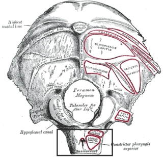
The pharyngeal tubercle is a part of the occipital bone of the head and neck. It is located on the lower surface of the basilar part of occipital bone. It is the site of attachment of the pharyngeal raphe.

The posterior auricular nerve is a nerve of the head. It is a branch of the facial nerve. It communicates with branches from the vagus nerve, the great auricular nerve, and the lesser occipital nerve. Its auricular branch supplies the posterior auricular muscle, the intrinsic muscles of the auricle, and gives sensation to the auricle. Its occipital branch supplies the occipitalis muscle.

