Corrugator supercilii muscle

Last updated
Corrugator supercilii muscle
Gray379corrugator.png
Anatomical right orbicularis oculi muscle (notice the corrugator muscle at the top)
Corrugator supercilii.png
Corrugator supercilii
Details
Origin Supraorbital ridge ( Superciliary arches )
Insertion Forehead skin, near eyebrow
Artery Ophthalmic artery
Nerve Facial nerve
Actions Wrinkles forehead
Identifiers
Latin musculus corrugator supercilii
TA98 A04.1.03.018
TA2 2071
FMA 46794
Anatomical terms of muscle

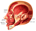
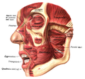
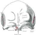
The corrugator supercilii muscle is a small, [1] narrow,[ citation needed ] pyramidal muscle [1] of the face.[ citation needed ] It arises from the medial end of the superciliary arch; it inserts into the deep surface of the skin of the eyebrow.

Contents

It draws the eyebrow downward and medially, producing the vertical "frowning" wrinkles of the forehead. It may be thought as the principal muscle in the facial expression of suffering. It also shields the eyes from strong sunlight.

Structure

The corrugator supercilii muscle is located at the medial end of the eyebrow. Its fibers pass laterally and somewhat superiorly from its origin to its insertion. [1]

Origin

It arises from bone at the medial extremity of the superciliary arch. [1]

Insertion

It inserts between the palpebral and orbital portions of the orbicularis oculi muscle. It inserts into the deep surface of the skin of the eyebrow, above the middle of the orbital arch.[ citation needed ]

Innervation

Motor innervation is provided by the temporal branches of facial nerve (CN VII). [1]

Vasculature

The muscle receives arterial supply from adjacent arteries - mostly from the superficial temporal artery, and the ophthalmic artery. [1]

Relations

It is situated deep to the frontalis muscle (of the occipitofrontalis muscle) and orbicularis oculi muscle, with which it partially blends.[ clarification needed ] [1] Its fibres are situated between the palpebral and orbital portions of the orbicularis oculi muscle.[ citation needed ]

The supratrochlear nerve passes between this muscle and the frontalis muscle. [2]

Function

The muscle acts in tandem with the orbicularis oculi muscle. The corrugator supercilii muscle acts upon the skin of the forehead superior to the middle of the supraorbital margin, [1] drawing the eyebrow inferomedially to produce vertical wrinkles of the forehead [3] just superior to the nose. [1] It is the "frowning" muscle, and may be regarded as the principal muscle in the expression of suffering. [4] It also contracts to prevent high sun glare, [4] pulling the eyebrows toward the bridge of the nose, making a roof over the area above the middle corner of the eye and typical forehead furrows [4] to shield the eye from excessively bright sunlight. [1]

Clinical significance

The muscle is sometimes surgically severed or paralysed with botulinum toxin as a preventive treatment for some types of migraine or for aesthetic reasons. [5]

Etymology

The name corrugator supercilii comes from Latin, and means wrinkler of the eyebrows.

Additional images

Related Research Articles

<span class="mw-page-title-main">Facial nerve</span> Cranial nerve VII, for the face and tasting

The facial nerve, also known as the seventh cranial nerve, cranial nerve VII, or simply CN VII, is a cranial nerve that emerges from the pons of the brainstem, controls the muscles of facial expression, and functions in the conveyance of taste sensations from the anterior two-thirds of the tongue. The nerve typically travels from the pons through the facial canal in the temporal bone and exits the skull at the stylomastoid foramen. It arises from the brainstem from an area posterior to the cranial nerve VI and anterior to cranial nerve VIII.

Articles related to anatomy include:

<span class="mw-page-title-main">Forehead</span> Area of the head between the eyebrows and the hairline

In human anatomy, the forehead is an area of the head bounded by three features, two of the skull and one of the scalp. The top of the forehead is marked by the hairline, the edge of the area where hair on the scalp grows. The bottom of the forehead is marked by the supraorbital ridge, the bone feature of the skull above the eyes. The two sides of the forehead are marked by the temporal ridge, a bone feature that links the supraorbital ridge to the coronal suture line and beyond. However, the eyebrows do not form part of the forehead.

<span class="mw-page-title-main">Supraorbital nerve</span> Branch of the frontal nerve

The supraorbital nerve is one of two branches of the frontal nerve, itself a branch of the ophthalmic nerve. The other branch of the frontal nerve is the supratrochlear nerve.

<span class="mw-page-title-main">Procerus muscle</span> Small pyramidal slip of muscle deep to the superior orbital nerve, artery and vein

The procerus muscle is a small pyramidal slip of muscle deep to the superior orbital nerve, artery and vein. Procerus is Latin, meaning tall or extended.

<span class="mw-page-title-main">Ophthalmic artery</span> Artery of the head

The ophthalmic artery (OA) is an artery of the head. It is the first branch of the internal carotid artery distal to the cavernous sinus. Branches of the ophthalmic artery supply all the structures in the orbit around the eye, as well as some structures in the nose, face, and meninges. Occlusion of the ophthalmic artery or its branches can produce sight-threatening conditions.

<span class="mw-page-title-main">Orbicularis oculi muscle</span> Facial muscle that closes the eyelids

The orbicularis oculi is a muscle in the face that closes the eyelids. It arises from the nasal part of the frontal bone, from the frontal process of the maxilla in front of the lacrimal groove, and from the anterior surface and borders of a short fibrous band, the medial palpebral ligament.

<span class="mw-page-title-main">Frontalis muscle</span>

The frontalis muscle is a muscle which covers parts of the forehead of the skull. Some sources consider the frontalis muscle to be a distinct muscle. However, Terminologia Anatomica currently classifies it as part of the occipitofrontalis muscle along with the occipitalis muscle.

<span class="mw-page-title-main">Depressor supercilii muscle</span> Muscle of the eye

The depressor supercilii is an eye muscle of the human body. The nature of this muscle is in some dispute. Few printed anatomies include it and many authorities consider it to be part of the orbicularis oculi muscle.

<span class="mw-page-title-main">Supratrochlear nerve</span> Nerve of the forehead

The supratrochlear nerve is a branch of the frontal nerve, itself a branch of the ophthalmic nerve (CN V1) from the trigeminal nerve (CN V). It provides sensory innervation to the skin of the forehead and the upper eyelid.

<span class="mw-page-title-main">Temporal fascia</span>

The temporal fascia is a fascia of the head that covers the temporalis muscle and structures situated superior to the zygomatic arch.

<span class="mw-page-title-main">Infraorbital nerve</span> Branch of the maxillary nerve supplying the face

The infraorbital nerve is a branch of the maxillary nerve. It arises in the pterygopalatine fossa. It passes through the inferior orbital fissure to enter the orbit. It travels through the orbit, then enters and traverses the infraorbital canal, exiting the canal at the infraorbital foramen to reach the face. It provides sensory innervation to the skin and mucous membranes around the middle of the face.

<span class="mw-page-title-main">Medial palpebral ligament</span> Ligament connecting the maxilla and the lacrimal bone to the eyelids

The medial palpebral ligament is a ligament of the face. It attaches to the frontal process of the maxilla, the lacrimal groove, and the tarsus of each eyelid. It has a superficial (anterior) and a deep (posterior) layer, with many surrounding attachments. It connects the medial canthus of each eyelid to the medial part of the orbit. It is a useful point of fixation during eyelid reconstructive surgery.

<span class="mw-page-title-main">Angular artery</span> Artery of the face

The angular artery is an artery of the face. It is the terminal part of the facial artery. It ascends to the medial angle of the eye's orbit. It is accompanied by the angular vein. It ends by anastomosing with the dorsal nasal branch of the ophthalmic artery. It supplies the lacrimal sac, the orbicularis oculi muscle, and the outer side of the nose.

<span class="mw-page-title-main">Medial palpebral arteries</span>

The medial palpebral arteries are arteries of the head that contribute arterial blood supply to the eyelids. They are derived from the ophthalmic artery; a single medial palpebral artery issues from the ophthalmic artery before splitting into a superior and an inferior medial palpebral artery, each supplying one eyelid.

<span class="mw-page-title-main">Supratrochlear artery</span>

The supratrochlear artery is one of the terminal branches of the ophthalmic artery. It arises within the orbit. It exits the orbit alongside the supratrochlear nerve. It contributes arterial supply to the skin, muscles and pericranium of the forehead.

<span class="mw-page-title-main">Temporal branches of the facial nerve</span>

The temporal branches of the facial nerve crosses the zygomatic arch to the temporal region, supplying the auriculares anterior and superior, and joining with the zygomaticotemporal branch of the maxillary nerve, and with the auriculotemporal branch of the mandibular nerve.

<span class="mw-page-title-main">Zygomatic branches of the facial nerve</span> Nerves of the face

The zygomatic branches of the facial nerve (malar branches) are nerves of the face. They run across the zygomatic bone to the lateral angle of the orbit. Here, they supply the orbicularis oculi muscle, and join with filaments from the lacrimal nerve and the zygomaticofacial branch of the maxillary nerve (CN V2).

<span class="mw-page-title-main">Facial muscles</span> Muscles of the face

The facial muscles are a group of striated skeletal muscles supplied by the facial nerve that, among other things, control facial expression. These muscles are also called mimetic muscles. They are only found in mammals, although they derive from neural crest cells found in all vertebrates. They are the only muscles that attach to the dermis.

<span class="mw-page-title-main">Outline of human anatomy</span> Overview of and topical guide to human anatomy

The following outline is provided as an overview of and topical guide to human anatomy:

References

PD-icon.svgThis article incorporates text in the public domain from page 907 of the 20th edition of Gray's Anatomy (1918)

  1. 1 2 3 4 5 6 7 8 9 10 Standring, Susan (2020). Gray's Anatomy: The Anatomical Basis of Clinical Practice (42th ed.). New York. p. 624. ISBN   978-0-7020-7707-4. OCLC   1201341621.{{cite book}}: CS1 maint: location missing publisher (link)
  2. Lasaosa, S. Santos; Pérez, M. L. Cuadrado; Peral, A. L. Guerrero; Villanueva, M. Huerta; Porta-Etessam, J.; Pozo-Rosich, P.; Pareja, J. A. (2017-06-01). "Consensus recommendations for anaesthetic peripheral nerve block". Neurología (English Edition). 32 (5): 316–330. doi: 10.1016/j.nrleng.2016.04.002 . ISSN   2173-5808.
  3. Cattaneo, Luigi; Pavesi, Giovanni (2014-01-01). "The facial motor system". Neuroscience & Biobehavioral Reviews . 38: 135–159. doi:10.1016/j.neubiorev.2013.11.002. ISSN   0149-7634. PMID   24239732. S2CID   207090623 via ScienceDirect.
  4. 1 2 3 "eye, human."Encyclopædia Britannica from Encyclopædia Britannica 2006 Ultimate Reference Suite DVD 2009
  5. de Ru JA, Schellekens PP, Lohuis PJ (2011). "Corrugator supercilii transection for headache emanating from the frontal region: a clinical evaluation of ten patients". J Neural Transm (Vienna). 118 (11): 1571–4. doi:10.1007/s00702-011-0654-1. PMID   21597942. S2CID   23829349.{{cite journal}}: CS1 maint: multiple names: authors list (link)