Articles related to anatomy include:

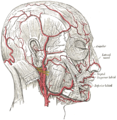
The external carotid artery is a major artery of the head and neck. It arises from the common carotid artery when it splits into the external and internal carotid artery. External carotid artery supplies blood to the face and neck.
The internal carotid artery is located in the inner side of the neck in contrast to the external carotid artery. In human anatomy, they arise from the common carotid arteries, where these bifurcate into the internal and external carotid arteries at cervical vertebrae C3 or C4; the internal carotid artery supplies the brain, including the eyes, while the external carotid nourishes other portions of the head, such as the face, the scalp, the skull, and the meninges.

The scalp is the anatomical area bordered by the human face at the front, and by the neck at the sides and back.
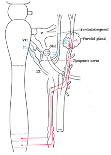
The auriculotemporal nerve is a branch of the mandibular nerve (CN V3) that runs with the superficial temporal artery and vein, and provides sensory innervation to various regions on the side of the head.

The temporal fossa is a fossa on the side of the skull bounded by the temporal lines and terminating below the level of the zygomatic arch.

The occipital artery arises from the external carotid artery opposite the facial artery. Its path is below the posterior belly of digastric to the occipital region. This artery supplies blood to the back of the scalp and sternocleidomastoid muscles, and deep muscles in the back and neck.

The superior thyroid artery arises from the external carotid artery just below the level of the greater cornu of the hyoid bone and ends in the thyroid gland.
The posterior auricular artery is a small artery that arises from the external carotid artery, above the digastric muscle and stylohyoid muscle, opposite the apex of the styloid process.

The maxillary artery supplies deep structures of the face. It branches from the external carotid artery just deep to the neck of the mandible.

The superficial temporal vein is a vein of the side of the head. It begins on the side and vertex of the skull in a network of veins which communicates with the frontal vein and supraorbital vein, with the corresponding vein of the opposite side, and with the posterior auricular vein and occipital vein. It ultimately crosses the posterior root of the zygomatic arch, enters the parotid gland, and unites with the internal maxillary vein to form the posterior facial vein.

The squamous part of temporal bone, or temporal squama, forms the front and upper part of the temporal bone, and is scale-like, thin, and translucent.

The middle cranial fossa, deeper than the anterior cranial fossa, is narrow medially and widens laterally to the sides of the skull. It is separated from the posterior fossa by the clivus and the petrous crest.

The deep cervical fascia lies under cover of the platysma, and invests the muscles of the neck; it also forms sheaths for the carotid vessels, and for the structures situated in front of the vertebral column. Its attachment to the hyoid bone prevents the formation of a dewlap.

The temporal fascia covers the temporalis muscle.
In anatomy, arterial tree is used to refer to all arteries and/or the branching pattern of the arteries. This article regards the human arterial tree. Starting from the aorta:

The submandibular triangle corresponds to the region of the neck immediately beneath the body of the mandible.

The carotid triangle is a portion of the anterior triangle of the neck.

The following outline is provided as an overview of and topical guide to human anatomy:
The cheek constitutes the facial periphery, plays a key role in the maintenance of oral competence and mastication, is involved in the facial manifestation of human emotion, and supports neighboring primary structures.

