Temporal bone

Last updated
Temporal bone
Temporal bone lateral5.png
Position of temporal bone (shown in green)
Temporal bone - animation 02.gif
Animation of the temporal bone
Details
Articulations Occipital, parietal, sphenoid, mandible and zygomatic
Identifiers
Latin os temporale
MeSH D013701
TA98 A02.1.06.001
TA2 641
FMA 52737
Anatomical terms of bone

The temporal bones are situated at the sides and base of the skull, and lateral to the temporal lobes of the cerebral cortex.

Contents

The temporal bones are overlaid by the sides of the head known as the temples, and house the structures of the ears. The lower seven cranial nerves and the major vessels to and from the brain traverse the temporal bone.

Structure

The temporal bone consists of four parts [1] [2] — the squamous, mastoid, petrous and tympanic parts. The squamous part is the largest and most superiorly positioned relative to the rest of the bone. The zygomatic process is a long, arched process projecting from the lower region of the squamous part and it articulates with the zygomatic bone. Posteroinferior to the squamous is the mastoid part. Fused with the squamous and mastoid parts and between the sphenoid and occipital bones lies the petrous part, which is shaped like a pyramid. The tympanic part is relatively small and lies inferior to the squamous part, anterior to the mastoid part, and superior to the styloid process. The styloid, from the Greek stylos, is a phallic shaped pillar directed inferiorly and anteromedially between the parotid gland and internal jugular vein. [3] An elongated or deviated styloid process can result from calcification of the stylohyoid ligament in a condition known as Eagle syndrome .

Borders

Development

The temporal bone is ossified from eight centers, exclusive of those for the internal ear and the tympanic ossicles: one for the squama including the zygomatic process, one for the tympanic part, four for the petrous and mastoid parts, and two for the styloid process. Just before the end of prenatal development [Fig. 6] the temporal bone consists of three principal parts:

  1. The squama is ossified in membrane from a single nucleus, which appears near the root of the zygomatic process about the second month.
  2. The petromastoid part is developed from four centers, which make their appearance in the cartilaginous ear capsule about the fifth or sixth month. One (proötic) appears in the neighborhood of the eminentia arcuata, spreads in front and above the internal auditory meatus and extends to the apex of the bone; it forms part of the cochlea, vestibule, superior semicircular canal, and medial wall of the tympanic cavity. A second (opisthotic) appears at the promontory on the medial wall of the tympanic cavity and surrounds the fenestra cochleæ; it forms the floor of the tympanic cavity and vestibule, surrounds the carotid canal, invests the lateral and lower part of the cochlea, and spreads medially below the internal auditory meatus. A third (pterotic) roofs in the tympanic cavity and antrum; while the fourth (epiotic) appears near the posterior semicircular canal and extends to form the mastoid process (Vrolik).
  3. The tympanic ring is an incomplete circle, in the concavity of which is a groove, the tympanic sulcus, for the attachment of the circumference of the eardrum (tympanic membrane). This ring expands to form the tympanic part, and is ossified in membrane from a single center which appears about the third month. The styloid process is developed from the proximal part of the cartilage of the second branchial or hyoid arch by two centers: one for the proximal part, the tympanohyal, appears before birth; the other, comprising the rest of the process, is named the stylohyal, and does not appear until after birth. The tympanic ring unites with the squama shortly before birth; the petromastoid part and squama join during the first year, and the tympanohyal portion of the styloid process about the same time [Fig. 7, 8]. The stylohyal does not unite with the rest of the bone until after puberty, and in some skulls never at all.

Postnatal development

Apart from size increase, the chief changes from birth through puberty in the temporal bone are as follows:

  1. The tympanic ring extends outward and backward to form the tympanic part. This extension does not, however, take place at an equal rate all around the circumference of the ring, but occurs more at its anterior and posterior portions. As these outgrowths meet, they create a foramen in the floor of the meatus, the foramen of Huschke. This foramen is usually closed about the fifth year, but may persist throughout life.
  2. The mandibular fossa is at first extremely shallow, and looks lateral and inferior; it deepens and directs more inferiorly over time. The part of the squama which forms the fossa lies at first below the level of the zygomatic process. As, the base of the skull thickens, this part of the squama is directed horizontal and inwards to contribute to the middle cranial fossa, and its surfaces look upward and downward; the attached portion of the zygomatic process everts and projects like a shelf at a right angle to the squama.
  3. The mastoid portion is at first flat, with the stylomastoid foramen and rudimentary styloid immediately behind the tympanic ring. With air cell development, the outer part of the mastoid component grows anteroinferiorly to form the mastoid process, with the styloid and stylomastoid foramen now on the under surface. The descent of the foramen is accompanied by a requisite lengthening of the facial canal.
  4. The downward and forward growth of the mastoid process also pushes forward the tympanic part; as a result, its portion that formed the original floor of the meatus, and contained the foramen of Huschke, rotates to become the anterior wall.
  5. The fossa subarcuata is nearly effaced.

Trauma

Temporal bone fractures were historically divided into three main categories, longitudinal, in which the vertical axis of the fracture paralleled the petrous ridge, horizontal, in which the axis of the fracture was perpendicular to the petrous ridge, and oblique, a mixed type with both longitudinal and horizontal components. Horizontal fractures were thought to be associated with injuries to the facial nerve, and longitudinal with injuries to the middle ear ossicles. [4] More recently, delineation based on disruption of the otic capsule has been found as more reliable in predicting complications such as facial nerve injury, sensorineural hearing loss, intracerebral hemorrhage, and cerebrospinal fluid otorrhea. [5]

Other animals

In many animals some of these parts stay separate through life:

In evolutionary terms, the temporal bone is derived from the fusion of many bones that are often separate in non-human mammals:

Etymology

Its exact etymology is unknown. [7] It is thought to be from the Old French temporal meaning "earthly", which is directly from the Latin tempus meaning "time, proper time or season." Temporal bones are situated on the sides of the skull, where grey hairs usually appear early on. Or it may relate to the pulsations of the underlying superficial temporal artery, marking the time we have left here. There is also a probable connection with the Greek verb temnion, to wound in battle. The skull is thin in this area and presents a vulnerable area for a blow from a battle axe. [8] Another possible etymology is described at Temple (anatomy).

Additional images

Pathology

Glomus jugulare tumor:

See also

Related Research Articles

<span class="mw-page-title-main">Facial nerve</span> Cranial nerve VII, for the face and tasting

The facial nerve, also known as the seventh cranial nerve, cranial nerve VII, or simply CN VII, is a cranial nerve that emerges from the pons of the brainstem, controls the muscles of facial expression, and functions in the conveyance of taste sensations from the anterior two-thirds of the tongue. The nerve typically travels from the pons through the facial canal in the temporal bone and exits the skull at the stylomastoid foramen. It arises from the brainstem from an area posterior to the cranial nerve VI and anterior to cranial nerve VIII.

Articles related to anatomy include:

<span class="mw-page-title-main">Glossopharyngeal nerve</span> Cranial nerve IX, for the tongue and pharynx

The glossopharyngeal nerve, also known as the ninth cranial nerve, cranial nerve IX, or simply CN IX, is a cranial nerve that exits the brainstem from the sides of the upper medulla, just anterior to the vagus nerve. Being a mixed nerve (sensorimotor), it carries afferent sensory and efferent motor information. The motor division of the glossopharyngeal nerve is derived from the basal plate of the embryonic medulla oblongata, whereas the sensory division originates from the cranial neural crest.

<span class="mw-page-title-main">Occipital bone</span> Bone of the neurocranium

The occipital bone is a cranial dermal bone and the main bone of the occiput. It is trapezoidal in shape and curved on itself like a shallow dish. The occipital bone overlies the occipital lobes of the cerebrum. At the base of the skull in the occipital bone, there is a large oval opening called the foramen magnum, which allows the passage of the spinal cord.

<span class="mw-page-title-main">Posterior cranial fossa</span> Area of the cranium containing the brainstem and cerebellum

The posterior cranial fossa is the part of the cranial cavity located between the foramen magnum, and tentorium cerebelli. It is formed by the sphenoid bones, temporal bones, and occipital bone. It lodges the cerebellum, and parts of the brainstem.

<span class="mw-page-title-main">Tympanic cavity</span> Small cavity surrounding the bones of the middle ear

The tympanic cavity is a small cavity surrounding the bones of the middle ear. Within it sit the ossicles, three small bones that transmit vibrations used in the detection of sound.
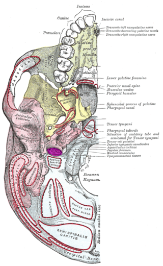
<span class="mw-page-title-main">Jugular fossa</span>

The jugular fossa is a deep depression in the inferior part of the temporal bone at the base of the skull. It lodges the bulb of the internal jugular vein.

<span class="mw-page-title-main">Internal auditory meatus</span> Canal within the temporal bone

The internal auditory meatus is a canal within the petrous part of the temporal bone of the skull between the posterior cranial fossa and the inner ear.

<span class="mw-page-title-main">Greater wing of sphenoid bone</span> Large part of the skull front behind the eye socket

The greater wing of the sphenoid bone, or alisphenoid, is a bony process of the sphenoid bone, positioned in the skull behind each eye. There is one on each side, extending from the side of the body of the sphenoid and curving upward, laterally, and backward.

<span class="mw-page-title-main">Transverse sinuses</span>

The transverse sinuses, within the human head, are two areas beneath the brain which allow blood to drain from the back of the head. They run laterally in a groove along the interior surface of the occipital bone. They drain from the confluence of sinuses to the sigmoid sinuses, which ultimately connect to the internal jugular vein. See diagram : labeled under the brain as "SIN. TRANS.".

<span class="mw-page-title-main">Squamous part of temporal bone</span> Front and upper part of the sides of the skull base

The squamous part of temporal bone, or temporal squama, forms the front and upper part of the temporal bone, and is scale-like, thin, and translucent.

<span class="mw-page-title-main">Mastoid part of the temporal bone</span> Back part of the sides of the skull base

The mastoid part of the temporal bone is the posterior (back) part of the temporal bone, one of the bones of the skull. Its rough surface gives attachment to various muscles and it has openings for blood vessels. From its borders, the mastoid part articulates with two other bones.

<span class="mw-page-title-main">Petrous part of the temporal bone</span> Feature at the base of the human skull

The petrous part of the temporal bone is pyramid-shaped and is wedged in at the base of the skull between the sphenoid and occipital bones. Directed medially, forward, and a little upward, it presents a base, an apex, three surfaces, and three angles, and houses in its interior, the components of the inner ear. The petrous portion is among the most basal elements of the skull and forms part of the endocranium. Petrous comes from the Latin word petrosus, meaning "stone-like, hard". It is one of the densest bones in the body. In other mammals, it is a separate bone, the petrosal bone.

<span class="mw-page-title-main">Tympanic part of the temporal bone</span> Middle part of the sides of the skull base, surrounding the ear canal

The tympanic part of the temporal bone is a curved plate of bone lying below the squamous part of the temporal bone, in front of the mastoid process, and surrounding the external part of the ear canal.

<span class="mw-page-title-main">Middle cranial fossa</span>

The middle cranial fossa is formed by the sphenoid bones, and the temporal bones. It lodges the temporal lobes, and the pituitary gland. It is deeper than the anterior cranial fossa, is narrow medially and widens laterally to the sides of the skull. It is separated from the posterior cranial fossa by the clivus and the petrous crest.

<span class="mw-page-title-main">Infratemporal fossa</span> Cavity that is part of the skull

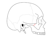
The infratemporal fossa is an irregularly shaped cavity that is a part of the skull. It is situated below and medial to the zygomatic arch. It is not fully enclosed by bone in all directions. It contains superficial muscles, including the lower part of the temporalis muscle, the lateral pterygoid muscle, and the medial pterygoid muscle. It also contains important blood vessels such as the middle meningeal artery, the pterygoid plexus, and the retromandibular vein, and nerves such as the mandibular nerve (CN V3) and its branches.

<span class="mw-page-title-main">Tympanic nerve</span> Branch of the glossopharyngeal nerve

The tympanic nerve is a branch of the glossopharyngeal nerve found near the ear. It gives sensation to the middle ear, the Eustachian tube, the parotid gland, and mastoid air cells. It gives parasympathetic to supply to the parotid gland via the otic ganglion and the auriculotemporal nerve.

<span class="mw-page-title-main">Zygomatic process</span>

The zygomatic processes are three processes (protrusions) from other bones of the skull which each articulate with the zygomatic bone. The three processes are:

<span class="mw-page-title-main">Mastoid cells</span> Air-filled cavities in the temporal bone

The mastoid cells are air-filled cavities within the mastoid process of the temporal bone of the cranium. The mastoid cells are a form of skeletal pneumaticity. Infection in these cells is called mastoiditis.

<span class="mw-page-title-main">Outline of human anatomy</span> Overview of and topical guide to human anatomy

The following outline is provided as an overview of and topical guide to human anatomy:

References

PD-icon.svgThis article incorporates text in the public domain from page 138 of the 20th edition of Gray's Anatomy (1918)

  1. "SKULL ANATOMY - TEMPORAL BONE". 28 June 2017.
  2. Temporal bone anatomy on CT 2012-12-22
  3. Chaurasia, BD (31 January 2013). Human Anatomy Volume 3 (Sixth ed.). CBS Publishers and Distributors Pvt Ltd. pp. 41–43. ISBN   9788123923321.
  4. Brodie, HA; Thompson, TC (March 1997). "Management of complications from 820 temporal bone fractures". The American Journal of Otology. 18 (2): 188–97. PMID   9093676.
  5. Little, SC; Kesser, BW (December 2006). "Radiographic classification of temporal bone fractures: clinical predictability using a new system". Archives of Otolaryngology–Head & Neck Surgery. 132 (12): 1300–4. doi: 10.1001/archotol.132.12.1300 . PMID   17178939.
  6. 1 2 Romer, Alfred Sherwood; Parsons, Thomas S. (1977). The Vertebrate Body. Colorado, PA: Holt-Saunders International. pp. XXX. ISBN   0-03-910284-X.
  7. "Temporal | Search Online Etymology Dictionary".
  8. "Etymology of Head Terms". www.dartmouth.edu. Archived from the original on 2008-05-09.
  9. "Glomus jugulare tumor: MedlinePlus Medical Encyclopedia". medlineplus.gov. Retrieved 2017-05-02.
  10. Sanei Taheri, Morteza; Zare Mehrjardi, Mohammad (2016-07-21). "Imaging of temporal bone lesions: developmental and inflammatory conditions".{{cite journal}}: Cite journal requires |journal= (help)