In anatomy, the zygomatic arch, or cheek bone, is a part of the skull formed by the zygomatic process of the temporal bone and the temporal process of the zygomatic bone, the two being united by an oblique suture ; the tendon of the temporal muscle passes medial to the arch, to gain insertion into the coronoid process of the mandible (jawbone).
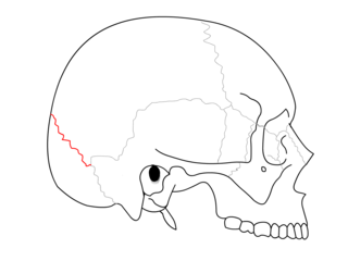
The lambdoid suture is a dense, fibrous connective tissue joint on the posterior aspect of the skull that connects the parietal bones with the occipital bone. It is continuous with the occipitomastoid suture.

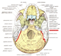
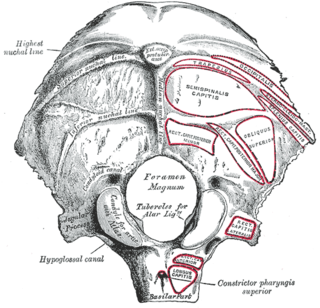
The longus capitis muscle, is broad and thick above, narrow below, and arises by four tendinous slips, from the anterior tubercles of the transverse processes of the third, fourth, fifth, and sixth cervical vertebræ, and ascends, converging toward its fellow of the opposite side, to be inserted into the inferior surface of the basilar part of the occipital bone.

The hypoglossal canal is a foramen in the occipital bone of the skull. It is hidden medially and superiorly to each occipital condyle. The hypoglossal nerve traverses the canal.

The jugular fossa is a deep depression in the inferior part of the base of the skull. More specifically, it is located in the temporal bone, posterior to the carotid canal and the aquæductus cochleæ. It is of variable depth and size in different skulls; it lodges the bulb of the internal jugular vein.
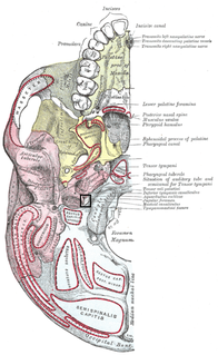
The jugular foramen is a large foramen (opening) in the base of the skull, located behind the carotid canal. It is formed in front by the petrous portion of the temporal bone, and behind by the occipital bone; it is generally larger on the right than on the left side.

The greater wing of the sphenoid bone, or alisphenoid, is a bony process of the sphenoid bone; there is one on each side, extending from the side of the body of the sphenoid and curving upward, laterally, and backward.

The mastoid part of the temporal bone is the posterior (back) part of the temporal bone, one of the bones of the skull. Its rough surface gives attachment to various muscles and it has openings for blood vessels. From its borders, the mastoid part articulates with two other bones.

The temporal styloid process is a process of bone that extends down from the temporal bone of the human skull, just below the ear.

In the lateral part of the jugular fossa of the temporal bone is the mastoid canaliculus for the entrance of the auricular branch of the vagus nerve.

The Sphenofrontal suture is the cranial suture between the sphenoid bone and the frontal bone.
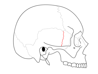
The Sphenosquamosal suture is a cranial suture between the sphenoid bone and the squama of the temporal bone.

The mastoid foramen is a hole in the posterior border of the temporal bone. It transmits a Mastoid emissary vein to the sigmoid sinus and a small branch of the occipital artery, the posterior meningeal artery to the dura mater.

This grooved surface of the foramen magnum is separated on either side from the petrous portion of the temporal bone by the petro-occipital fissure, which is occupied in the fresh state by a plate of cartilage; the fissure is continuous behind with the jugular foramen, and its margins are grooved for the inferior petrosal sinus.
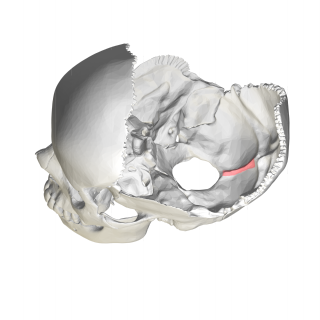
The condylar canal is a canal in the condyloid fossa of the lateral parts of occipital bone behind the occipital condyle. Resection of the rectus capitis posterior major and minor muscles reveals the bony recess leading to the condylar canal, which is situated posterior and lateral to the occipital condyle. It is immediately superior to the extradural vertebral artery, which makes a loop above the posterior C1 ring to enter the foramen magnum. The anteriomedial wall of the condylar canal thickens to join the foramen magnum rim and connect to the occipital condyle.
The upper surface of the lateral parts of occipital bone presents an oval eminence, the jugular tubercle, which overlies the hypoglossal canal and is sometimes crossed by an oblique groove for the glossopharyngeal, vagus, and accessory nerves.

Along the internal surface of the occipital bone, at the point of intersection of the four divisions of the cruciform eminence is the internal occipital protuberance. Running transversely on either side is a groove for the transverse sinus.
In the occipital bone, the lower division of the cruciate eminence is prominent, and is named the internal occipital crest; it bifurcates near the foramen magnum and gives attachment to the falx cerebelli; in the attached margin of this falx is the occipital sinus, which is sometimes duplicated.
The great wings, or alae-sphenoids, are two strong processes of bone, which arise from the sides of the body, and are curved upward, lateralward, and backward; the posterior part of each projects as a triangular process which fits into the angle between the squama and the petrous portion of the temporal bone and presents at its apex a downwardly directed process, the spina angularis. It serves as the origin for the sphenomandibular ligament.

Along the internal surface of the occipital bone, running laterally between the superior and inferior fossae of the cruciform eminence is the groove for transverse sinus. The transverse sinuses travel along this groove.