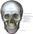
The frontal bone or sincipital bone is a bone in the human skull. The bone consists of two portions. These are the vertically oriented squamous part, and the horizontally oriented orbital part, making up the bony part of the forehead, part of the bony orbital cavity holding the eye, and part of the bony part of the nose respectively. The name comes from the Latin word frons.

In anatomy, the zygomatic arch, or cheek bone, is a part of the skull formed by the zygomatic process of the temporal bone and the temporal process of the zygomatic bone, the two being united by an oblique suture ; the tendon of the temporal muscle passes medial to the arch, to gain insertion into the coronoid process of the mandible (jawbone).

The orbital or horizontal part of the frontal bone consists of two thin triangular plates, the orbital plates, which form the vaults of the orbits, and are separated from one another by a median gap, the ethmoidal notch.
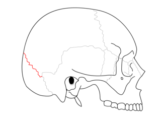
The lambdoid suture is a dense, fibrous connective tissue joint on the posterior aspect of the skull that connects the parietal bones with the occipital bone. It is continuous with the occipitomastoid suture.

The coronal suture is a dense, fibrous connective tissue joint that separates the two parietal bones from the frontal bone of the skull.

The condyloid process or condylar process is the process on the human and other mammalian species' mandibles that ends in a condyle, the mandibular condyle. It is thicker than the coronoid process of the mandible and consists of two portions: the condyle and the constricted portion which supports it, the neck.

The greater wing of the sphenoid bone, or alisphenoid, is a bony process of the sphenoid bone, positioned in the skull behind each eye. There is one on each side, extending from the side of the body of the sphenoid and curving upward, laterally, and backward.
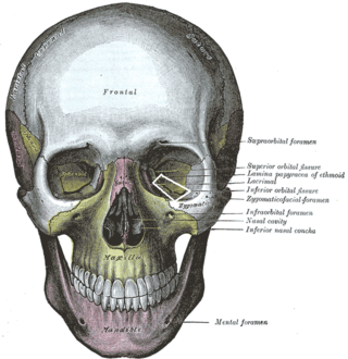
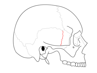
The inferior orbital fissure is a gap between the greater wing of sphenoid bone, and the maxilla. It connects the orbit (anteriorly) with the infratemporal fossa and pterygopalatine fossa (posteriorly).
The squamous part of the frontal bone is the superior portion when viewed in standard anatomical orientation. There are two surfaces of the squamous part of the frontal bone: the external surface, and the internal surface.

The squamosal suture, or squamous suture, arches backward from the pterion and connects the temporal squama with the lower border of the parietal bone: this suture is continuous behind with the short, nearly horizontal parietomastoid suture, which unites the mastoid process of the temporal with the region of the mastoid angle of the parietal bone. The term parietotemporal suture may refer to both of these sutures or exclusively to the parietomastoid suture and its use is, therefore, best avoided.

The sphenozygomatic suture is the cranial suture between the sphenoid bone and the zygomatic bone.

The sphenofrontal suture is the cranial suture between the sphenoid bone and the frontal bone.

The zygomaticotemporal suture is the cranial suture between the zygomatic bone and the temporal bone. This is part of the zygomatic arch. Movement at the suture decreases with development during aging. It has a complex internal structure.

The occipitomastoid suture or occipitotemporal suture is the cranial suture between the occipital bone and the mastoid portion of the temporal bone.
The sphenosquamosal suture is a cranial suture between the sphenoid bone and the squama of the temporal bone.

The anterior nasal spine, or anterior nasal spine of maxilla, is a bony projection in the skull that serves as a cephalometric landmark. The anterior nasal spine is the projection formed by the fusion of the two maxillary bones at the intermaxillary suture. It is placed at the level of the nostrils, at the uppermost part of the philtrum. It rarely fractures.

The zygomatic processes are three processes (protrusions) from other bones of the skull which each articulate with the zygomatic bone. The three processes are:
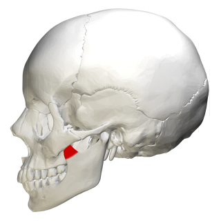
In human anatomy, the mandible's coronoid process is a thin, triangular eminence, which is flattened from side to side and varies in shape and size. Its anterior border is convex and is continuous below with the anterior border of the ramus. Its posterior border is concave and forms the anterior boundary of the mandibular notch. The lateral surface is smooth, and affords insertion to the temporalis and masseter muscles. Its medial surface gives insertion to the temporalis, and presents a ridge which begins near the apex of the process and runs downward and forward to the inner side of the last molar tooth.
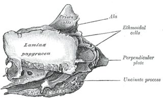
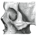
The orbital lamina of ethmoid bone is a smooth, oblong, paper-thin bone plate which forms the lateral wall of the labyrinth of the ethmoid bone. It covers the middle and posterior ethmoidal cells, and forms a large part of the medial wall of the orbit.
Koerner's septum is an anatomic boundary in the temporal bone formed by the petrosquamous suture between the petrous and squamosal portions of the mastoid air cells, at the anatomic level of the mastoid antrum. Along with the middle ear ossicles, it is usually eroded in middle ear cholesteatomas. Superiorly, this continues as the petrosquamous suture, a normal anatomic structure that can be mistaken for fractures on temporal bone CT. It is surgically important as it may cause difficulty in locating the antrum and the deeper cells and thus may lead to incomplete removal of disease at mastoidectomy.