Base of skull

Last updated
Skull base
Skull interior anatomy.svg
Base of the skull, inferior or inner surface
Skull base anatomy.jpg
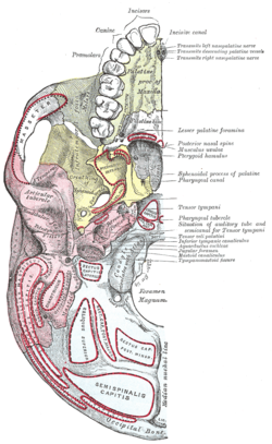
Base of the skull, exterior or outer surface. Showing various muscle attachments.
Details
Identifiers
Latin basis cranii
externa et. interna
MeSH D019291
TA98 A02.1.00.044
TA2 447
FMA 52801
Anatomical terms of bone

The base of skull, also known as the cranial base or the cranial floor, is the most inferior area of the skull. It is composed of the endocranium and the lower parts of the calvaria.

Contents

Structure

Base of the skull. Inferior surface, attachment of muscles marked in red. Gray187.png
Base of the skull. Inferior surface, attachment of muscles marked in red.

Structures found at the base of the skull are for example:

Bones

There are five bones that make up the base of the skull:

Exobasis Exobasis.jpg
Exobasis

Sinuses

Foramina of the skull

Endobasis-resistances beams Endobasis - resistances beams.jpg
Endobasis-resistances beams
Endobasis-resistances nodes Endobasis - resistances nodes.jpg
Endobasis-resistances nodes

Sutures

Other

The foramina in the base of the skull are exit and entry points for veins, arteries and cranial nerves. Skull brain human normal.svg
The foramina in the base of the skull are exit and entry points for veins, arteries and cranial nerves.
The cranial nerves as they exit through various foramina. Skull and brainstem inner ear.svg
The cranial nerves as they exit through various foramina.

Development

During the fetal period, the geometry of the cranial base and its fossae: anterior, middle and posterior undergoes rapid changes. The anterior part of the cranial base undergoes changes rapidly particularly in the first trimester, and cranial defects can frequently develop during this period. Growth of the anterior part of cranial base is uneven during the prenatal period. Allometric growth is observed in the first trimester, with the longitudinal dimension increasing from 5 to 17 millimeters between the 8th and 14th weeks of fetal life. Simultaneously, the angle of the anterior cranial fossa decreases, and its depth increases toward the middle fossa. In the second trimester, growth continues but becomes more uniform, and changes in the angle of the anterior fossa are minor. The angle between the lesser wings of the sphenoid bone gradually decreases with increasing depth of the anterior fossa in the frontal plane. [1]

Additional images

  1. Kedzia, Alicja; Derkowski, Wojciech; Glonek, Michal (2006). "The structure and function of the human skull based on studies using computer image analysis methods in the fetal period.". Computer-aided scientific research =: Komputerowe wspomaganie badań naukowych (in Polish and English). Vol. 13. Wrocław: Wrocław Scientific Society = Wrocławskie Towarzystwo Naukowe. Wydawnictwo. pp. 241–246. ISBN   978-83-7374-043-3.