Foramen lacerum

Last updated
Foramen lacerum
Gray193.png
Base of the skull. Upper surface. (Foramen lacerum is labeled at center left, and is visible as the large hole between yellow sphenoid, red temporal, and blue occipital)
Details
System skeletal
Parts temporal bone, sphenoid bone, occipital bone
Identifiers
Latin foramen lacerum
TA98 A02.1.00.055
TA2 459
FMA 54809
Anatomical terminology

The foramen lacerum (Latin : lacerated piercing) is a triangular hole in the base of skull. It is located between the sphenoid bone, the apex of the petrous part of the temporal bone, and the basilar part of the occipital bone.

Contents

Structure

The foramen lacerum (Latin : lacerated piercing) is a triangular hole in the base of skull. It is located between 3 bones:

It is the junction point of 3 sutures of the skull:

Contents

Structures passing through the foramen lacerum include:

Relations

It is situated anteromedially to the carotid canal. [1] :776

The internal carotid artery passes from the carotid canal in the base of the skull, emerging and coursing superior to foramen lacerum as it exits the carotid canal; the internal carotid artery does not travel through foramen lacerum (the segment of the internal carotid artery that travels superior to the foramen lacerum is called the lacerum segment). [7]

Development

The foramen lacerum fills with cartilage after birth. [1] :776

Clinical significance

The foramen lacerum has been described as a portal of entry into the cranium for tumours, including nasopharyngeal carcinoma, juvenile angiofibroma, adenoid cystic carcinoma, malignant melanoma, and lymphoma. [8] [9]

History

The first recorded mention of the foramen lacerum was by anatomist Wenzel Gruber in 1869. [10] [8] Study of the foramen has been neglected for many years because of the small role it plays in intracranial surgery. [8]

Additional images

Related Research Articles

Articles related to anatomy include:

<span class="mw-page-title-main">Foramen ovale (skull)</span> Hole in the sphenoid bone of the skull

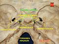
The foramen ovale is a hole in the posterior part of the sphenoid bone, posterolateral to the foramen rotundum. It is one of the larger of the several holes in the skull. It transmits the mandibular nerve, a branch of the trigeminal nerve.

<span class="mw-page-title-main">Sphenoid bone</span> Bone at the front of the skull

The sphenoid bone is an unpaired bone of the neurocranium. It is situated in the middle of the skull towards the front, in front of the basilar part of the occipital bone. The sphenoid bone is one of the seven bones that articulate to form the orbit. Its shape somewhat resembles that of a butterfly or bat with its wings extended.

<span class="mw-page-title-main">Occipital bone</span> Skull bone, lower back of cranium

The occipital bone is a cranial dermal bone and the main bone of the occiput. It is trapezoidal in shape and curved on itself like a shallow dish. The occipital bone overlies the occipital lobes of the cerebrum. At the base of the skull in the occipital bone, there is a large oval opening called the foramen magnum, which allows the passage of the spinal cord.

<span class="mw-page-title-main">Internal carotid artery</span> Artery of the human brain

The internal carotid artery is an artery in the neck which supplies the anterior circulation of the brain.

<span class="mw-page-title-main">Posterior cranial fossa</span> Area of the cranium containing the brainstem and cerebellum

The posterior cranial fossa is the part of the cranial cavity located between the foramen magnum, and tentorium cerebelli. It is formed by the sphenoid bones, temporal bones, and occipital bone. It lodges the cerebellum, and parts of the brainstem.

<span class="mw-page-title-main">Cavernous sinus</span> Sinus in the human head

The cavernous sinus within the human head is one of the dural venous sinuses creating a cavity called the lateral sellar compartment bordered by the temporal bone of the skull and the sphenoid bone, lateral to the sella turcica.

<span class="mw-page-title-main">Foramen spinosum</span> Hole in the sphenoid bone of the skull

The foramen spinosum is a small open hole in the greater wing of the sphenoid bone that gives passage to the middle meningeal artery and vein, and the meningeal branch of the mandibular nerve.

<span class="mw-page-title-main">Jugular foramen</span> Opening in the base of the skull allowing many structures to pass

A jugular foramen is one of the two large foramina (openings) in the base of the skull, located behind the carotid canal. It is formed by the temporal bone and the occipital bone. It allows many structures to pass, including the inferior petrosal sinus, three cranial nerves, the sigmoid sinus, and meningeal arteries.

<span class="mw-page-title-main">Greater wing of sphenoid bone</span> Large part of the skull front behind the eye socket

The greater wing of the sphenoid bone, or alisphenoid, is a bony process of the sphenoid bone, positioned in the skull behind each eye. There is one on each side, extending from the side of the body of the sphenoid and curving upward, laterally, and backward.

<span class="mw-page-title-main">Carotid canal</span> Hole in the temporal bone in the skull for the internal carotid artery to enter the skull

The carotid canal is a passage in the petrous part of the temporal bone of the skull through which the internal carotid artery and its internal carotid (nervous) plexus pass from the neck into the cranial cavity.

<span class="mw-page-title-main">Petrous part of the temporal bone</span> Feature at the base of the human skull

The petrous part of the temporal bone is pyramid-shaped and is wedged in at the base of the skull between the sphenoid and occipital bones. Directed medially, forward, and a little upward, it presents a base, an apex, three surfaces, and three angles, and houses in its interior, the components of the inner ear. The petrous portion is among the most basal elements of the skull and forms part of the endocranium. Petrous comes from the Latin word petrosus, meaning "stone-like, hard". It is one of the densest bones in the body. In other mammals, it is a separate bone, the petrosal bone.

<span class="mw-page-title-main">Basilar part of occipital bone</span> Section of the main skull bone that extends forward and upward

The basilar part of the occipital bone extends forward and upward from the foramen magnum, and presents in front an area more or less quadrilateral in outline.

<span class="mw-page-title-main">Middle cranial fossa</span>

The middle cranial fossa is formed by the sphenoid bones, and the temporal bones. It lodges the temporal lobes, and the pituitary gland. It is deeper than the anterior cranial fossa, is narrow medially and widens laterally to the sides of the skull. It is separated from the posterior cranial fossa by the clivus and the petrous crest.

<span class="mw-page-title-main">Infratemporal fossa</span> Cavity that is part of the skull

The infratemporal fossa is an irregularly shaped cavity that is a part of the skull. It is situated below and medial to the zygomatic arch. It is not fully enclosed by bone in all directions. It contains superficial muscles, including the lower part of the temporalis muscle, the lateral pterygoid muscle, and the medial pterygoid muscle. It also contains important blood vessels such as the middle meningeal artery, the pterygoid plexus, and the retromandibular vein, and nerves such as the mandibular nerve (CN V3) and its branches.

<span class="mw-page-title-main">Nerve of pterygoid canal</span>

The nerve of the pterygoid canal is formed by the union of the (parasympathetic) greater petrosal nerve and (sympathetic) deep petrosal nerve within the cartilaginous substance filling the foramen lacerum. From the foramen lacerum, the nerve of the pterygoid canal passes through the pterygoid canal to reach the pterygopalatine fossa, ending at the pterygopalatine ganglion.

<span class="mw-page-title-main">Body of sphenoid bone</span>

The body of the sphenoid bone, more or less cubical in shape, is hollowed out in its interior to form two large cavities, the sphenoidal sinuses, which are separated from each other by a septum.

<span class="mw-page-title-main">Clivus (anatomy)</span> Bony part of the skull base

The clivus or Blumenbach clivus is a bony part of the cranium at the base of the skull. It is a shallow depression behind the dorsum sellae of the sphenoid bone. It slopes gradually to the anterior part of the basilar occipital bone at its junction with the sphenoid bone. It extends to the foramen magnum. It is related to the pons and the abducens nerve.

<span class="mw-page-title-main">Base of skull</span> Inferior area of the skull, composed of the endocranium and lower parts of the skull roof

The base of skull, also known as the cranial base or the cranial floor, is the most inferior area of the skull. It is composed of the endocranium and the lower parts of the calvaria.

<span class="mw-page-title-main">Outline of human anatomy</span> Overview of and topical guide to human anatomy

The following outline is provided as an overview of and topical guide to human anatomy:

References

  1. 1 2 3 4 5 6 7 Drake, Richard L.; Vogl, Wayne; Tibbitts, Adam W.M. Mitchell; illustrations by Richard; Richardson, Paul (2005). Gray's anatomy for students. Philadelphia: Elsevier/Churchill Livingstone. ISBN   978-0-8089-2306-0.
  2. Chae, Ricky; Rubio, Roberto Rodriguez (2020). "14 - Anatomy of petrous face". Handbook of Clinical Neurology. Vol. 170. Elsevier. pp. 143–156. doi:10.1016/B978-0-12-822198-3.00036-7. ISBN   978-0-12-822198-3. ISSN   0072-9752. PMID   32586486. S2CID   220077819.
  3. 1 2 Netter, Frank H.; Norton, Neil S. (2011). Netter's Head and Neck Anatomy for Dentistry (2nd ed.). Elsevier, Saunders. pp. 47–50.
  4. Goosmann, Madeline M.; Dalvin, Mark (2023), "Anatomy, Head and Neck, Deep Petrosal Nerve", StatPearls, Treasure Island (FL): StatPearls Publishing, PMID   30521238 , retrieved 2023-07-31
  5. Seker, Askin; Martins, Carolina; Rhoton, Albert L. (2010). "2 - Meningeal Anatomy". Meningiomas. Saunders. pp. 11–51. doi:10.1016/B978-1-4160-5654-6.00002-7. ISBN   978-1-4160-5654-6.{{cite book}}: CS1 maint: location missing publisher (link)
  6. Sinnatamby, Chummy S. (2011). Last's Anatomy (12th ed.). Elsevier Australia. p. 364. ISBN   978-0-7295-3752-0.
  7. Tubbs, R. Shane; Shoja, Mohammadali M.; Loukas, Marios (25 April 2016). Bergman's Comprehensive Encyclopedia of Human Anatomic Variation. John Wiley & Sons. p. 450. ISBN   9781118430279.
  8. 1 2 3 Tauber, M; van Loveren, HR; Jallo, G; Romano, A; Keller, JT (February 1999). "The enigmatic foramen lacerum". Neurosurgery. 44 (2): 386–91, discussion 391-3. doi:10.1097/00006123-199902000-00083. PMID   9932893.
  9. Christodouleas, Boris Hristov, Steven H. Lin, John P. (2010). Radiation oncology : a question-based review. Philadelphia, Pa.: Lippincott Williams & Wilkins. p. 138. ISBN   978-1608314447.{{cite book}}: CS1 maint: multiple names: authors list (link)
  10. Gruber, Wenzel (1869). Beitrage Zur Anatomie Des Schadelgrundes. ISBN   9781162306223.