Posterior cranial fossa

Last updated
Posterior cranial fossa
Posterior cranial fossa boundaries.svg
Gray193 - Cranial fossae.png
Base of the skull. Upper surface. The Posterior cranial fossa is colored in blue, yellow, and red.
Details
Identifiers
Latin fossa cranii posterior
MeSH D003388
TA98 A02.1.00.050
TA2 453
FMA 54368
Anatomical terminology

The posterior cranial fossa is the part of the cranial cavity located between the foramen magnum, and tentorium cerebelli. It is formed by the sphenoid bones, temporal bones, and occipital bone. It lodges the cerebellum, and parts of the brainstem.

Anatomy

The posterior cranial fossa is formed by the sphenoid bones, temporal bones, and occipital bone. [1] It is the most inferior of the fossae.[ citation needed ] It houses the cerebellum, medulla oblongata, and pons. [2]

Boundaries

Anteriorly, the posterior cranial fossa is bounded by the dorsum sellae, posterior aspect of the body of sphenoid bone, and the basilar part of occipital bone/clivus. [2]

Laterally, it is bounded by the petrous parts and mastoid parts of the temporal bones, and the lateral parts of occipital bone. [2]

Posteriorly, it is bounded by the squamous part of occipital bone. [2]

Features

Foramen magnum

The foramen magnum is a large opening of the floor of the posterior cranial fossa, its most conspicuous feature. [2]

Internal acoustic meatus

Lies in the anterior wall of the posterior cranial fossa. It transmits the facial (VII) and vestibulocochlear (VIII) cranial nerves into a canal in the petrous temporal bone.

Jugular foramen

Lies between the inferior edge of the petrous temporal bone and the adjacent occipital bone and transmits the internal jugular vein (actually begins here), the glossopharyngeal nerve (CN IX), vagus nerve (CN X), and accessory nerve (CN XI).

Hypoglossal canal

Lies at the anterolateral margins of the foramen magnum and transmits the hypoglossal nerve (CN XII).

Other

Also visible in the posterior cranial fossa are depressions caused by the venous sinuses returning blood from the brain to the venous circulation: Right and left transverse sinuses which meet at the confluence of sinuses (marked by the internal occipital protuberance).

The transverse sinuses pass horizontally from the most posterior point of the occiput.

Where the apex of the petrous temporal meets the squamous temporal, the transverse sinuses lead into sigmoid (S-shaped) sinuses (one on each side).

These pass along the articulation between the posterior edge of the petrous temporal bone and the anterior edge of the occipital bones to the jugular foramen, where the sigmoid sinus becomes the internal jugular vein.

Note that a superior petrosal sinus enters the junction of the transverse and sigmoid sinuses. Also an inferior petrosal sinus enters the sigmoid sinus near the jugular foramen.

The posterior cranial fossa is formed in the endocranium, and holds the most basal parts of the brain.

Clinical significance

An underdeveloped posterior cranial fossa can cause Arnold–Chiari malformation. These can be either acquired or congenital disorders.[ citation needed ]

Additional images

See also

Related Research Articles

Articles related to anatomy include:

<span class="mw-page-title-main">Glossopharyngeal nerve</span> Cranial nerve IX, for the tongue and pharynx

The glossopharyngeal nerve, also known as the ninth cranial nerve, cranial nerve IX, or simply CN IX, is a cranial nerve that exits the brainstem from the sides of the upper medulla, just anterior to the vagus nerve. Being a mixed nerve (sensorimotor), it carries afferent sensory and efferent motor information. The motor division of the glossopharyngeal nerve is derived from the basal plate of the embryonic medulla oblongata, whereas the sensory division originates from the cranial neural crest.

<span class="mw-page-title-main">Occipital bone</span> Skull bone, lower back of cranium

The occipital bone is a cranial dermal bone and the main bone of the occiput. It is trapezoidal in shape and curved on itself like a shallow dish. The occipital bone overlies the occipital lobes of the cerebrum. At the base of the skull in the occipital bone, there is a large oval opening called the foramen magnum, which allows the passage of the spinal cord.

<span class="mw-page-title-main">Temporal bone</span> Sides and base of skull, connecting to the jaw and occipital bone

The temporal bones are situated at the sides and base of the skull, and lateral to the temporal lobes of the cerebral cortex.

<span class="mw-page-title-main">Internal carotid artery</span> Artery of the human brain

The internal carotid artery is an artery in the neck which supplies the anterior circulation of the brain.

<span class="mw-page-title-main">Cavernous sinus</span> Sinus in the human head

The cavernous sinus within the human head is one of the dural venous sinuses creating a cavity called the lateral sellar compartment bordered by the temporal bone of the skull and the sphenoid bone, lateral to the sella turcica.

<span class="mw-page-title-main">Inferior petrosal sinus</span>

The inferior petrosal sinuses are two small sinuses situated on the inferior border of the petrous part of the temporal bone, one on each side. Each inferior petrosal sinus drains the cavernous sinus into the internal jugular vein.

<span class="mw-page-title-main">Jugular foramen</span> Opening in the base of the skull allowing many structures to pass

A jugular foramen is one of the two large foramina (openings) in the base of the skull, located behind the carotid canal. It is formed by the temporal bone and the occipital bone. It allows many structures to pass, including the inferior petrosal sinus, three cranial nerves, the sigmoid sinus, and meningeal arteries.

<span class="mw-page-title-main">Greater wing of sphenoid bone</span> Large part of the skull front behind the eye socket

The greater wing of the sphenoid bone, or alisphenoid, is a bony process of the sphenoid bone, positioned in the skull behind each eye. There is one on each side, extending from the side of the body of the sphenoid and curving upward, laterally, and backward.

<span class="mw-page-title-main">Sigmoid sinus</span>

The sigmoid sinuses, also known as the pars sigmoid, are paired dural venous sinuses within the skull that receive blood from posterior transverse sinuses.

<span class="mw-page-title-main">Transverse sinuses</span>

The transverse sinuses, within the human head, are two areas beneath the brain which allow blood to drain from the back of the head. They run laterally in a groove along the interior surface of the occipital bone. They drain from the confluence of sinuses to the sigmoid sinuses, which ultimately connect to the internal jugular vein. See diagram : labeled under the brain as "SIN. TRANS.".
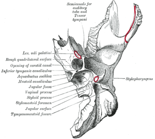
<span class="mw-page-title-main">Carotid canal</span> Hole in the temporal bone in the skull for the internal carotid artery to enter the skull

The carotid canal is a passage in the petrous part of the temporal bone of the skull through which the internal carotid artery and its internal carotid (nervous) plexus pass from the neck into the cranial cavity.

<span class="mw-page-title-main">Petrous part of the temporal bone</span> Feature at the base of the human skull

The petrous part of the temporal bone is pyramid-shaped and is wedged in at the base of the skull between the sphenoid and occipital bones. Directed medially, forward, and a little upward, it presents a base, an apex, three surfaces, and three angles, and houses in its interior, the components of the inner ear. The petrous portion is among the most basal elements of the skull and forms part of the endocranium. Petrous comes from the Latin word petrosus, meaning "stone-like, hard". It is one of the densest bones in the body. In other mammals, it is a separate bone, the petrosal bone.

<span class="mw-page-title-main">Lateral parts of occipital bone</span>

The lateral parts of the occipital bone are situated at the sides of the foramen magnum; on their under surfaces are the condyles for articulation with the superior facets of the atlas.

<span class="mw-page-title-main">Middle cranial fossa</span>

The middle cranial fossa is formed by the sphenoid bones, and the temporal bones. It lodges the temporal lobes, and the pituitary gland. It is deeper than the anterior cranial fossa, is narrow medially and widens laterally to the sides of the skull. It is separated from the posterior cranial fossa by the clivus and the petrous crest.

<span class="mw-page-title-main">Mastoid cells</span> Air-filled cavities in the temporal bone

The mastoid cells are air-filled cavities within the mastoid process of the temporal bone of the cranium. The mastoid cells are a form of skeletal pneumaticity. Infection in these cells is called mastoiditis.

<span class="mw-page-title-main">Body of sphenoid bone</span>

The body of the sphenoid bone, more or less cubical in shape, is hollowed out in its interior to form two large cavities, the sphenoidal sinuses, which are separated from each other by a septum.

<span class="mw-page-title-main">Base of skull</span> Inferior area of the skull, composed of the endocranium and lower parts of the skull roof

The base of skull, also known as the cranial base or the cranial floor, is the most inferior area of the skull. It is composed of the endocranium and the lower parts of the calvaria.

<span class="mw-page-title-main">Outline of human anatomy</span> Overview of and topical guide to human anatomy

The following outline is provided as an overview of and topical guide to human anatomy:

<span class="mw-page-title-main">Hiatus for lesser petrosal nerve</span> Opening in the temporal bone

The hiatus for lesser petrosal nerve is a hiatus in the petrous part of the temporal bone which transmits the lesser petrosal nerve. It is located posterior to the groove for the superior petrosal sinus and posterolateral to the jugular foramen.

References

  1. Rea, Paul (2016). "3 - Neck". Essential Clinically Applied Anatomy of the Peripheral Nervous System in the Head and Neck. Academic Press. pp. 131–183. doi:10.1016/B978-0-12-803633-4.00003-X. ISBN   978-0-12-803633-4.
  2. 1 2 3 4 5 Rea, Paul (2016). "3 - Neck". Essential Clinically Applied Anatomy of the Peripheral Nervous System in the Head and Neck. Academic Press. pp. 131–183. doi:10.1016/B978-0-12-803633-4.00003-X. ISBN   978-0-12-803633-4.