The facial nerve is the seventh cranial nerve, or simply CN VII. It emerges from the pons of the brainstem, controls the muscles of facial expression, and functions in the conveyance of taste sensations from the anterior two-thirds of the tongue. The nerves typically travels from the pons through the facial canal in the temporal bone and exits the skull at the stylomastoid foramen. It arises from the brainstem from an area posterior to the cranial nerve VI and anterior to cranial nerve VIII.
Articles related to anatomy include:

The glossopharyngeal nerve, known as the ninth cranial nerve, is a mixed nerve that carries afferent sensory and efferent motor information. It exits the brainstem out from the sides of the upper medulla, just anterior to the vagus nerve. The motor division of the glossopharyngeal nerve is derived from the basal plate of the embryonic medulla oblongata, while the sensory division originates from the cranial neural crest.

The accessory nerve is a cranial nerve that supplies the sternocleidomastoid and trapezius muscles. It is considered as the eleventh of twelve pairs of cranial nerves, or simply cranial nerve XI, as part of it was formerly believed to originate in the brain. The sternocleidomastoid muscle tilts and rotates the head, while the trapezius muscle, connecting to the scapula, acts to shrug the shoulder.

At the base of the skull, the foramen ovale is one of the larger of the several holes that transmit nerves through the skull. The foramen ovale is situated in the posterior part of the sphenoid bone, posterolateral to the foramen rotundum.

The internal carotid artery is located in the inner side of the neck in contrast to the external carotid artery. In human anatomy, they arise from the common carotid arteries where these bifurcate into the internal and external carotid arteries at cervical vertebral level 3 or 4; the internal carotid artery supplies the brain including eyes, while the external carotid nourishes other portions of the head, such as the face, scalp, skull, and meninges.

The posterior cranial fossa is part of the cranial cavity, located between the foramen magnum and tentorium cerebelli. It contains the brainstem and cerebellum.

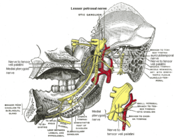
The otic ganglion is a small parasympathetic ganglion located immediately below the foramen ovale in the infratemporal fossa and on the medial surface of the mandibular nerve. It is functionally associated with the glossopharyngeal nerve and innervates the parotid gland for salivation.

The tympanic cavity is a small cavity surrounding the bones of the middle ear. Within it sit the ossicles, three small bones that transmit vibrations used in the detection of sound.
Petrosal nerve may refer to:
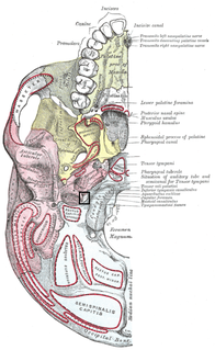
The jugular foramen is a large foramen (opening) in the base of the skull, located behind the carotid canal. It is formed in front by the petrous portion of the temporal bone, and behind by the occipital bone; it is generally larger on the right than on the left side.

The petrous part of the temporal bone is pyramid-shaped and is wedged in at the base of the skull between the sphenoid and occipital bones. Directed medially, forward, and a little upward, it presents a base, an apex, three surfaces, and three angles, and houses in its interior, the components of the inner ear. The petrous portion is among the most basal elements of the skull and forms part of the endocranium. Petrous comes from the Latin word petrosus, meaning "stone-like, hard". It is one of the densest bones in the body.

The lateral parts of the occipital bone are situated at the sides of the foramen magnum; on their under surfaces are the condyles for articulation with the superior facets of the atlas.
The inferior ganglion of the glossopharyngeal nerve is a sensory ganglion. It is larger than and below the superior ganglion of the glossopharyngeal nerve. It is located within the jugular foramen.

The middle cranial fossa, deeper than the anterior cranial fossa, is narrow medially and widens laterally to the sides of the skull. It is separated from the posterior fossa by the clivus and the petrous crest.
The tympanic nerve is a branch of the glossopharyngeal nerve found near the ear.
The lesser petrosal nerve is the general visceral efferent (GVE) component of the glossopharyngeal nerve, carrying parasympathetic preganglionic fibers from the tympanic plexus to the parotid gland. It synapses in the otic ganglion, from where the postganglionic fibers emerge.
The superior ganglion of the glossopharyngeal nerve is a sensory ganglion of the peripheral nervous system. It is located within the jugular foramen where the glossopharyngeal nerve exits the skull. It is smaller than and above the inferior ganglion of the glossopharyngeal nerve.

The base of skull, also known as the cranial base or the cranial floor, is the most inferior area of the skull. It is composed of the endocranium and the lower parts of the skull roof.

The following outline is provided as an overview of and topical guide to human anatomy: