Carotid canal

Last updated
Carotid canal
Gray141.png
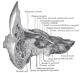
Left temporal bone. Inferior surface. ("Opening of carotid canal" labeled at center left.)
Details
Part of temporal bone
System skeletal
Identifiers
Latin canalis caroticus
TA98 A02.1.06.013
TA2 651
FMA 55805
Anatomical terms of bone

The carotid canal is a passage in the petrous part of the temporal bone of the skull through which the internal carotid artery and its internal carotid (nervous) plexus pass from the neck into (the middle cranial fossa of) the cranial cavity.

Contents

Structure

The carotid canal is located within the middle cranial fossa, at the petrous part of the temporal bone. Anteriorly, it is limited by posterior margin of the greater wing of sphenoid bone. Posteromedially, it is limited by basilar part of occipital bone. It is divided in three parts, namely, ascending petrous, transverse petrous, and ascending cavernous parts. The carotid canal has two openings, namely internal and external openings. The internal opening is situated laterally to foramen lacerum. The external opening of the carotid canal is located posterolaterally to the foramen lacerum. Both internal and external openings of the carotid canal lies anterior to the jugular foramen, where the latter is located inside the posterior cranial fossa. [1] The carotid canal is separated from middle ear and inner ear by a thin plate of bone. [2]

Contents

The canal transmits internal carotid artery together with its associated nervous plexus and venous plexus. [1]

Clinical significance

Any skull fractures that damage the carotid canal can put the internal carotid artery at risk. [3] Angiography can be used to ensure that there is no damage, and to aid in treatment if there is. [3]

Other animals

The carotid canal starts on the inferior surface of the temporal bone of the skull at the external opening of the carotid canal (also referred to as the carotid foramen). The canal ascends at first superiorly, and then, making a bend, runs anteromedially. Its internal opening is near the foramen lacerum, above which the internal carotid artery passes on its way anteriorly to the cavernous sinus. [4]

The carotid canal allows the internal carotid artery to pass into the cranium, [4] [5] as well as the carotid plexus traveling on the artery. [4]

The carotid plexus contains sympathetics to the head from the superior cervical ganglion. [4] They have several motor functions: raise the eyelid (superior tarsal muscle), dilate pupil (pupillary dilator muscle), innervate sweat glands of face and scalp and constricts blood vessels in the head.

Additional images

Related Research Articles

Articles related to anatomy include:

<span class="mw-page-title-main">Glossopharyngeal nerve</span> Cranial nerve IX, for the tongue and pharynx

The glossopharyngeal nerve, also known as the ninth cranial nerve, cranial nerve IX, or simply CN IX, is a cranial nerve that exits the brainstem from the sides of the upper medulla, just anterior to the vagus nerve. Being a mixed nerve (sensorimotor), it carries afferent sensory and efferent motor information. The motor division of the glossopharyngeal nerve is derived from the basal plate of the embryonic medulla oblongata, whereas the sensory division originates from the cranial neural crest.

<span class="mw-page-title-main">Occipital bone</span> Skull bone, lower back of cranium

The occipital bone is a cranial dermal bone and the main bone of the occiput. It is trapezoidal in shape and curved on itself like a shallow dish. The occipital bone overlies the occipital lobes of the cerebrum. At the base of the skull in the occipital bone, there is a large oval opening called the foramen magnum, which allows the passage of the spinal cord.

<span class="mw-page-title-main">Temporal bone</span> Sides and base of skull, connecting to the jaw and occipital bone

The temporal bones are situated at the sides and base of the skull, and lateral to the temporal lobes of the cerebral cortex.

<span class="mw-page-title-main">Internal carotid artery</span> Artery of the human brain

The internal carotid artery is an artery in the neck which supplies the anterior circulation of the brain.

<span class="mw-page-title-main">Foramen lacerum</span> Triangular hole in the base of the skull

The foramen lacerum is a triangular hole in the base of skull. It is located between the sphenoid bone, the apex of the petrous part of the temporal bone, and the basilar part of the occipital bone.

<span class="mw-page-title-main">Posterior cranial fossa</span> Area of the cranium containing the brainstem and cerebellum

The posterior cranial fossa is the part of the cranial cavity located between the foramen magnum, and tentorium cerebelli. It is formed by the sphenoid bones, temporal bones, and occipital bone. It lodges the cerebellum, and parts of the brainstem.

<span class="mw-page-title-main">Tympanic cavity</span> Small cavity surrounding the bones of the middle ear

The tympanic cavity is a small cavity surrounding the bones of the middle ear. Within it sit the ossicles, three small bones that transmit vibrations used in the detection of sound.

<span class="mw-page-title-main">Greater petrosal nerve</span> Nerve in the skull

The greater petrosal nerve is a nerve of the head mainly containing pre-ganglionic parasympathetic fibres which ultimately synapse in the pterygopalatine ganglion. It branches from the facial nerve and is derived from the parasympathetic part of the nervus intermedius component of CN VII, with its cell bodies located in the superior salivary nucleus. In the connective tissue substance of the foramen lacerum, the greater petrosal nerve unites with the (sympathetic) deep petrosal nerve to form the nerve of the pterygoid canal which proceeds to the pterygopalatine ganglion.

<span class="mw-page-title-main">Cavernous sinus</span> Sinus in the human head

The cavernous sinus within the human head is one of the dural venous sinuses creating a cavity called the lateral sellar compartment bordered by the temporal bone of the skull and the sphenoid bone, lateral to the sella turcica.

<span class="mw-page-title-main">Internal auditory meatus</span> Canal within the temporal bone

The internal auditory meatus is a canal within the petrous part of the temporal bone of the skull between the posterior cranial fossa and the inner ear.

<span class="mw-page-title-main">Greater wing of sphenoid bone</span> Large part of the skull front behind the eye socket

The greater wing of the sphenoid bone, or alisphenoid, is a bony process of the sphenoid bone; there is one on each side, extending from the side of the body of the sphenoid and curving upward, laterally, and backward.

<span class="mw-page-title-main">Trigeminal ganglion</span> Sensory ganglion of the trigeminal nerve

The trigeminal ganglion is the sensory ganglion of each trigeminal nerve. The trigeminal ganglion is located within the trigeminal cave, a cavity formed by dura mater.

<span class="mw-page-title-main">Petrous part of the temporal bone</span> Feature at the base of the human skull

The petrous part of the temporal bone is pyramid-shaped and is wedged in at the base of the skull between the sphenoid and occipital bones. Directed medially, forward, and a little upward, it presents a base, an apex, three surfaces, and three angles, and houses in its interior, the components of the inner ear. The petrous portion is among the most basal elements of the skull and forms part of the endocranium. Petrous comes from the Latin word petrosus, meaning "stone-like, hard". It is one of the densest bones in the body. In other mammals, it is a separate bone, the petrosal bone.

<span class="mw-page-title-main">Middle cranial fossa</span>

The middle cranial fossa is formed by the sphenoid bones, and the temporal bones. It lodges the temporal lobes, and the pituitary gland. It is deeper than the anterior cranial fossa, is narrow medially and widens laterally to the sides of the skull. It is separated from the posterior cranial fossa by the clivus and the petrous crest.

<span class="mw-page-title-main">Infratemporal fossa</span> Cavity that is part of the skull

The infratemporal fossa is an irregularly shaped cavity that is a part of the skull. It is situated below and medial to the zygomatic arch. It is not fully enclosed by bone in all directions. It contains superficial muscles, including the lower part of the temporalis muscle, the lateral pterygoid muscle, and the medial pterygoid muscle. It also contains important blood vessels such as the middle meningeal artery, the pterygoid plexus, and the retromandibular vein, and nerves such as the mandibular nerve (CN V3) and its branches.

<span class="mw-page-title-main">Mastoid cells</span> Air-filled cavities in the temporal bone

The mastoid cells are air-filled cavities within the mastoid process of the temporal bone of the cranium. The mastoid cells are a form of skeletal pneumaticity. Infection in these cells is called mastoiditis.

<span class="mw-page-title-main">Base of skull</span> Inferior area of the skull, composed of the endocranium and lower parts of the skull roof

The base of skull, also known as the cranial base or the cranial floor, is the most inferior area of the skull. It is composed of the endocranium and the lower parts of the calvaria.

<span class="mw-page-title-main">Outline of human anatomy</span> Overview of and topical guide to human anatomy

The following outline is provided as an overview of and topical guide to human anatomy:

<span class="mw-page-title-main">Parapharyngeal space</span>

The parapharyngeal space, is a potential space in the head and the neck. It has clinical importance in otolaryngology due to parapharyngeal space tumours and parapharyngeal abscess developing in this area. It is also a key anatomic landmark for localizing disease processes in the surrounding spaces of the neck; the direction of its displacement indirectly reflects the site of origin for masses or infection in adjacent areas, and consequently their appropriate differential diagnosis.

References

PD-icon.svgThis article incorporates text in the public domain from page 143 of the 20th edition of Gray's Anatomy (1918)

  1. 1 2 Naidoo N, Lazarus L, Ajayi NO, Satyapal KS (2017). "An anatomical investigation of the carotid canal". Folia Morphologica. 76 (2): 289–294. doi: 10.5603/FM.a2016.0060 . PMID   27714731.
  2. Ryan, Stephanie (2011). "2". Anatomy for diagnostic imaging (Third ed.). Elsevier Ltd. p. 80. ISBN   9780702029714.
  3. 1 2 Houseman, Clifford M.; Belverud, Shawn A.; Narayan, Raj K. (2012). "20 - Closed Head Injury". Principles of Neurological Surgery (3rd ed.). Saunders. pp. 325–347. doi:10.1016/C2009-0-52989-3. ISBN   978-1-4377-0701-4.
  4. 1 2 3 4 Kumar, Amarendhra M.; Roman-Auerhahn, Margo Ruth (2005-01-01). "1 - Anatomy of the Canine and Feline Ear". Small Animal Ear Diseases (2nd ed.). Saunders. pp. 1–21. doi:10.1016/B0-72-160137-5/50004-0. ISBN   978-0-7216-0137-3.{{cite book}}: CS1 maint: date and year (link)
  5. Maynard, Robert Lewis; Downes, Noel (2019-01-01). "7 - The Cardiovascular System". Anatomy and Histology of the Laboratory Rat in Toxicology and Biomedical Research. Academic Press. pp. 77–90. doi:10.1016/B978-0-12-811837-5.00007-1. ISBN   978-0-12-811837-5.{{cite book}}: CS1 maint: date and year (link)