Perpendicular plate of ethmoid bone

Last updated
Perpendicular plate of ethmoid bone
Gray150.png
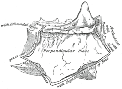
Perpendicular plate of ethmoid. Shown by removing the right labyrinth.
Gray151.png
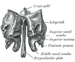
Ethmoid bone from behind.
Details
Identifiers
Latin lamina perpendicularis ossis ethmoidalis
TA A02.1.07.006
FMA 52891
Anatomical terms of bone

The perpendicular plate of the ethmoid bone (vertical plate) is a thin, flattened lamina, polygonal in form, which descends from the under surface of the cribriform plate, and assists in forming the septum of the nose; it is generally deflected a little to one or other side. The anterior border articulates with the spine of the frontal bone and the crest of the nasal bones.

Ethmoid bone bone in the skull that separates the nasal cavity from the brain

The ethmoid bone is an unpaired bone in the skull that separates the nasal cavity from the brain. It is located at the roof of the nose, between the two orbits. The cubical bone is lightweight due to a spongy construction. The ethmoid bone is one of the bones that make up the orbit of the eye.

In elementary geometry, a polygon is a plane figure that is described by a finite number of straight line segments connected to form a closed polygonal chain or polygonal circuit. The solid plane region, the bounding circuit, or the two together, may be called a polygon.

Cribriform plate

In human anatomy, the cribriform plate of the ethmoid bone is received into the ethmoidal notch of the frontal bone and roofs in the nasal cavities.

Contents

The posterior border articulates by its upper half with the sphenoidal crest, by its lower with the vomer.

Vomer facial bone

The vomer is one of the unpaired facial bones of the skull. It is located in the midsagittal line, and articulates with the sphenoid, the ethmoid, the left and right palatine bones, and the left and right maxillary bones. The vomer forms the inferior part of the nasal septum, with the superior part formed by the perpendicular plate of the ethmoid bone. The name is derived from the Latin word for a ploughshare and the shape of the bone.

The inferior border is thicker than the posterior, and serves for the attachment of the septal nasal cartilage of the nose.

Septal nasal cartilage

The septal nasal cartilage is composed of hyaline cartilage. It is somewhat quadrilateral in form, thicker at its margins than at its center, and completes the separation between the nasal cavities in front.

The surfaces of the plate are smooth, except above, where numerous grooves and canals are seen; these lead from the medial foramina on the cribriform plate and lodge filaments of the olfactory nerves.

Additional images

Related Research Articles

Sphenoid bone unpaired bone situated at the front middle of the skull in front of the temporal bone and basilar part of the occipital bone

The sphenoid bone is an unpaired bone of the neurocranium. It is situated in the middle of the skull towards the front, in front of the temporal bone and the basilar part of the occipital bone. The sphenoid bone is one of the seven bones that articulate to form the orbit. Its shape somewhat resembles that of a butterfly or bat with its wings extended.

Lacrimal bone smallest and most fragile bone of the human skull and face

The lacrimal bone is a small and fragile bone of the facial skeleton; it is roughly the size of the little fingernail. It is situated at the front part of the medial wall of the orbit. It has two surfaces and four borders. Several bony landmarks of the lacrimal bone function in the process of lacrimation or crying. Specifically, the lacrimal bone helps form the nasolacrimal canal necessary for tear translocation. A depression on the anterior inferior portion of the bone, the lacrimal fossa, houses the membranous lacrimal sac. Tears or lacrimal fluid, from the lacrimal glands, collect in this sac during excessive lacrimation. The fluid then flows through the nasolacrimal duct and into the nasopharynx. This drainage results in what is commonly referred to a runny nose during excessive crying or tear production. Injury or fracture of the lacrimal bone can result in posttraumatic obstruction of the lacrimal pathways.

Inferior nasal concha facial bone

The inferior nasal concha is one of the three paired nasal conchae in the nose. It extends horizontally along the lateral wall of the nasal cavity and consists of a lamina of spongy bone, curled upon itself like a scroll,. The inferior nasal conchae are considered a pair of facial bones. As the air passes through the turbinates, the air is churned against these mucosa-lined bones in order to receive warmth, moisture and cleansing. Superior to inferior nasal concha are the middle nasal concha and superior nasal concha which arise from the cranial portion of the skull. Hence, these two are considered as a part of the cranial bones.

Nasal cavity

The nasal cavity is a large, air-filled space above and behind the nose in the middle of the face. The nasal septum divides the cavity into two fossae. Each fossa is the continuation of one of the two nostrils. The nasal cavity is the uppermost part of the respiratory system and provides the nasal passage for inhaled air from the nostrils to the nasopharynx and rest of the respiratory tract.

Nasal septum separator of the left and right airways in the nose

The nasal septum separates the left and right airways in the nose, dividing the two nostrils.

Orbital part of frontal bone

The orbital or horizontal part of the frontal bone consists of two thin triangular plates, the orbital plates, which form the vaults of the orbits, and are separated from one another by a median gap, the ethmoidal notch.

Sphenoidal conchae

The sphenoidal conchae are two thin, curved plates, situated at the anterior and lower part of the body of the sphenoid. An aperture of variable size exists in the anterior wall of each, and through this the sphenoidal sinus opens into the nasal cavity.

Ethmoidal labyrinth

The ethmoidal labyrinth or lateral mass of the ethmoid bone consists of a number of thin-walled cellular cavities, the ethmoid air cells, arranged in three groups, anterior, middle, and posterior, and interposed between two vertical plates of bone; the lateral plate forms part of the orbit, the medial plate forms part of the nasal cavity. In the disarticulated bone many of these cells are opened into, but when the bones are articulated, they are closed in at every part, except where they open into the nasal cavity.

Middle nasal concha

The medial surface of the labyrinth of ethmoid consists of a thin lamella, which descends from the under surface of the cribriform plate, and ends below in a free, convoluted margin, the middle nasal concha.

Squamous part of the frontal bone

There are two surfaces of the squamous part of the frontal bone: the external surface, and the internal surface.

Perpendicular plate of palatine bone

The perpendicular plate of palatine bone is the vertical part of the palatine bone, and is thin, of an oblong form, and presents two surfaces and four borders.

Horizontal plate of palatine bone

The horizontal plate of palatine bone is a quadrilateral part of the palatine bone, and has two surfaces and four borders.

Frontal process of maxilla

The frontal process of maxilla is a strong plate, which projects upward, medialward, and backward from the maxilla, forming part of the lateral boundary of the nose.

Body of sphenoid bone

The body of the sphenoid bone, more or less cubical in shape, is hollowed out in its interior to form two large cavities, the sphenoidal sinuses, which are separated from each other by a septum.

Human nose feature of the face

The human nose is the most protruding part of the face that bears the nostrils and is the first organ of the respiratory system. The nose is also the principal organ in the olfactory system. The shape of the nose is determined by the nasal bones and the nasal cartilages, including the nasal septum which separates the nostrils and divides the nasal cavity into two. On average the nose of a male is larger than that of a female.

Plate (anatomy)

A plate in animal anatomy may refer to several things:

References

This article incorporates text in the public domain from page 154 of the 20th edition of Gray's Anatomy (1918)

The public domain consists of all the creative works to which no exclusive intellectual property rights apply. Those rights may have expired, been forfeited, expressly waived, or may be inapplicable.

<i>Grays Anatomy</i> English-language textbook of human anatomy

Gray's Anatomy is an English language textbook of human anatomy originally written by Henry Gray and illustrated by Henry Vandyke Carter. Earlier editions were called Anatomy: Descriptive and Surgical, Anatomy of the Human Body and Gray's Anatomy: Descriptive and Applied, but the book's name is commonly shortened to, and later editions are titled, Gray's Anatomy. The book is widely regarded as an extremely influential work on the subject, and has continued to be revised and republished from its initial publication in 1858 to the present day. The latest edition of the book, the 41st, was published in September 2015.