Greater wing of sphenoid bone

Last updated
Greater wing of sphenoid bone
Gray145.png
Figure 1: Sphenoid bone, upper surface.
Gray146.png
Figure 2: Sphenoid bone, anterior and inferior surfaces.
Details
Identifiers
Latin ala major ossis sphenoidalis
TA98 A02.1.05.024
TA2 610
FMA 52868
Anatomical terms of bone

The greater wing of the sphenoid bone, or alisphenoid, is a bony process of the sphenoid bone, positioned in the skull behind each eye. There is one on each side, extending from the side of the body of the sphenoid and curving upward, laterally, and backward.

Contents

Structure

The greater wings of the sphenoid are two strong processes of bone, which arise from the sides of the body, and are curved upward, laterally, and backward; the posterior part of each projects as a triangular process that fits into the angle between the squamous and the petrous part of the temporal bone and presents at its apex a downward-directed process, the spine of sphenoid bone.

Cerebral surface

The superior or cerebral surface of each greater wing [Fig. 1] forms part of the middle cranial fossa; it is deeply concave, and presents depressions for the convolutions of the temporal lobe of the brain. It has a number of foramina (holes) in it:

Lateral surface

The lateral surface [Fig. 2] is convex, and divided by a transverse ridge, the infratemporal crest, into two portions.

It is pierced by the foramen ovale and foramen spinosum, and at its posterior part is the sphenoidal spine, which is frequently grooved on its medial surface for the chorda tympani nerve.

To the sphenoidal spine are attached the sphenomandibular ligament and the tensor veli palatini muscle.

Medial to the anterior extremity of the infratemporal crest is a triangular process that serves to increase the attachment of the lateral pterygoid muscle; extending downward and medialward from this process on to the front part of the lateral pterygoid plate is a ridge that forms the anterior limit of the infratemporal surface, and, in the articulated skull, the posterior boundary of the pterygomaxillary fissure.

Orbital surface

The orbital surface of the great wing [Fig. 2], smooth, and quadrilateral in shape, is directed forward and medially and forms the posterior part of the lateral wall of the orbit.

Margin

Commencing from behind [Fig. 2], that portion of the circumference of the great wing that extends from the body to the spine is irregular.

In front of the spine the circumference presents a concave, serrated edge, bevelled at the expense of the inner table below, and of the outer table above, for articulation with the squamous part of the temporal bone.

At the tip of the great wing is a triangular portion, bevelled at the expense of the internal surface, for articulation with the sphenoidal angle of the parietal bone; this region is named the pterion.

Medial to this is a triangular, serrated surface, for articulation with the frontal bone; this surface is continuous medially with the sharp edge that forms the lower boundary of the superior orbital fissure, and laterally with the serrated margin for articulation with the zygomatic bone.

Development

The greater wing of the sphenoid bone starts as a separate bone, and is still separate at birth in humans.

Function

The sphenoid bone assists with the formation of the base and the sides of the skull, and the floors and walls of the orbits. It is the site of attachment for most of the muscles of mastication. Many foramina and fissures are located in the sphenoid that carry nerves and blood vessels of the head and neck, such as the superior orbital fissure (with ophthalmic nerve), foramen rotundum (with maxillary nerve) and foramen ovale (with mandibular nerve). [1]

Clinical significance

In patients with neurofibromatosis type 1, malformation of the sphenoid bone wings may occur, due to aberrant cell development. This can ultimately lead to blindness if left untreated.

In other animals

In many mammals, e.g. the dog, the greater wing of the sphenoid bone stays through life a separate bone called the alisphenoid.

Additional images

Related Research Articles

Articles related to anatomy include:

<span class="mw-page-title-main">Zygomatic bone</span> Facial bone

In the human skull, the zygomatic bone, also called cheekbone or malar bone, is a paired irregular bone which articulates with the maxilla, the temporal bone, the sphenoid bone and the frontal bone. It is situated at the upper and lateral part of the face and forms the prominence of the cheek, part of the lateral wall and floor of the orbit, and parts of the temporal fossa and the infratemporal fossa. It presents a malar and a temporal surface; four processes, and four borders.

<span class="mw-page-title-main">Foramen ovale (skull)</span> Hole in the sphenoid bone of the skull

The foramen ovale is a hole in the posterior part of the sphenoid bone, posterolateral to the foramen rotundum. It is one of the larger of the several holes in the skull. It transmits the mandibular nerve, a branch of the trigeminal nerve.

<span class="mw-page-title-main">Sphenoid bone</span> Bone at the front of the skull

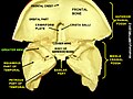
The sphenoid bone is an unpaired bone of the neurocranium. It is situated in the middle of the skull towards the front, in front of the basilar part of the occipital bone. The sphenoid bone is one of the seven bones that articulate to form the orbit. Its shape somewhat resembles that of a butterfly or bat with its wings extended.

<span class="mw-page-title-main">Palatine bone</span> Bone of the facial skeleton

In anatomy, the palatine bones are two irregular bones of the facial skeleton in many animal species, located above the uvula in the throat. Together with the maxillae, they comprise the hard palate.

<span class="mw-page-title-main">Orbit (anatomy)</span> Cavity or socket of the skull in which the eye and its appendages are situated

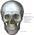
In anatomy, the orbit is the cavity or socket/hole of the skull in which the eye and its appendages are situated. "Orbit" can refer to the bony socket, or it can also be used to imply the contents. In the adult human, the volume of the orbit is 30 millilitres, of which the eye occupies 6.5 ml. The orbital contents comprise the eye, the orbital and retrobulbar fascia, extraocular muscles, cranial nerves II, III, IV, V, and VI, blood vessels, fat, the lacrimal gland with its sac and duct, the eyelids, medial and lateral palpebral ligaments, cheek ligaments, the suspensory ligament, septum, ciliary ganglion and short ciliary nerves.

<span class="mw-page-title-main">Middle meningeal artery</span> Artery supplying the meninges

The middle meningeal artery is typically the third branch of the first portion of the maxillary artery. After branching off the maxillary artery in the infratemporal fossa, it runs through the foramen spinosum to supply the dura mater and the calvaria. The middle meningeal artery is the largest of the three (paired) arteries that supply the meninges, the others being the anterior meningeal artery and the posterior meningeal artery.

<span class="mw-page-title-main">Cavernous sinus</span> Sinus in the human head

The cavernous sinus within the human head is one of the dural venous sinuses creating a cavity called the lateral sellar compartment bordered by the temporal bone of the skull and the sphenoid bone, lateral to the sella turcica.

<span class="mw-page-title-main">Foramen spinosum</span> Hole in the sphenoid bone of the skull

The foramen spinosum is a small open hole in the greater wing of the sphenoid bone that gives passage to the middle meningeal artery and vein, and the meningeal branch of the mandibular nerve.

<span class="mw-page-title-main">Pterygopalatine fossa</span> Fossa in the skull

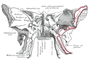
In human anatomy, the pterygopalatine fossa is a fossa in the skull. A human skull contains two pterygopalatine fossae—one on the left side, and another on the right side. Each fossa is a cone-shaped paired depression deep to the infratemporal fossa and posterior to the maxilla on each side of the skull, located between the pterygoid process and the maxillary tuberosity close to the apex of the orbit. It is the indented area medial to the pterygomaxillary fissure leading into the sphenopalatine foramen. It communicates with the nasal and oral cavities, infratemporal fossa, orbit, pharynx, and middle cranial fossa through eight foramina.

<span class="mw-page-title-main">Pterygoid processes of the sphenoid</span> Bone plates projecting from the sphenoid bone of the skull

The pterygoid processes of the sphenoid, one on either side, descend perpendicularly from the regions where the body and the greater wings of the sphenoid bone unite.

<span class="mw-page-title-main">Squamous part of temporal bone</span> Front and upper part of the sides of the skull base

The squamous part of temporal bone, or temporal squama, forms the front and upper part of the temporal bone, and is scale-like, thin, and translucent.

<span class="mw-page-title-main">Middle cranial fossa</span>

The middle cranial fossa is formed by the sphenoid bones, and the temporal bones. It lodges the temporal lobes, and the pituitary gland. It is deeper than the anterior cranial fossa, is narrow medially and widens laterally to the sides of the skull. It is separated from the posterior cranial fossa by the clivus and the petrous crest.

<span class="mw-page-title-main">Sphenoidal emissary foramen</span>

In the base of the skull, in the great wings of the sphenoid bone, medial to the foramen ovale, a small aperture, the sphenoidal emissary foramen, may occasionally be seen opposite the root of the pterygoid process. When present, it opens below near the scaphoid fossa. Vesalius was the first to describe and illustrate this foramen, and is also called the foramen Vesalius. Other names include foramen venosum and canaliculus sphenoidalis.

<span class="mw-page-title-main">Infratemporal fossa</span> Cavity that is part of the skull

The infratemporal fossa is an irregularly shaped cavity that is a part of the skull. It is situated below and medial to the zygomatic arch. It is not fully enclosed by bone in all directions. It contains superficial muscles, including the lower part of the temporalis muscle, the lateral pterygoid muscle, and the medial pterygoid muscle. It also contains important blood vessels such as the middle meningeal artery, the pterygoid plexus, and the retromandibular vein, and nerves such as the mandibular nerve (CN V3) and its branches.

<span class="mw-page-title-main">Sphenoidal process of palatine bone</span> Thin, compressed plate

The sphenoidal process of palatine bone is a thin, superomedially directed plate of bone. It is smaller and more inferior compared to the orbital process of palatine bone.

<span class="mw-page-title-main">Perpendicular plate of palatine bone</span>

The perpendicular plate of palatine bone is the vertical part of the palatine bone, and is thin, of an oblong form, and presents two surfaces and four borders.

<span class="mw-page-title-main">Body of sphenoid bone</span>

The body of the sphenoid bone, more or less cubical in shape, is hollowed out in its interior to form two large cavities, the sphenoidal sinuses, which are separated from each other by a septum.

<span class="mw-page-title-main">Base of skull</span> Inferior area of the skull, composed of the endocranium and lower parts of the skull roof

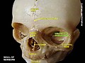
The base of skull, also known as the cranial base or the cranial floor, is the most inferior area of the skull. It is composed of the endocranium and the lower parts of the calvaria.

<span class="mw-page-title-main">Outline of human anatomy</span> Overview of and topical guide to human anatomy

The following outline is provided as an overview of and topical guide to human anatomy:

References

PD-icon.svgThis article incorporates text in the public domain from page 149 of the 20th edition of Gray's Anatomy (1918)

  1. Fehrenbach; Herring (2012). Illustrated Anatomy of the Head and Neck. Elsevier. p. 52. ISBN   978-1-4377-2419-6.