| Mandibular nerve | |
|---|---|
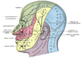
 Mandibular division of the trigeminal nerve. | |
 Mandibular division of trigeminal nerve, seen from the middle line. The small figure is an enlarged view of the otic ganglion. | |
| Details | |
| From | Trigeminal nerve (CN V) |
| Identifiers | |
| Latin | nervus mandibularis |
| MeSH | D008340 |
| TA98 | A14.2.01.064 |
| TA2 | 6246 |
| FMA | 52996 |
| Anatomical terms of neuroanatomy | |
In neuroanatomy, the mandibular nerve (V3) is the largest of the three divisions of the trigeminal nerve, the fifth cranial nerve (CN V). Unlike the other divisions of the trigeminal nerve (ophthalmic nerve, maxillary nerve) which contain only afferent fibers, the mandibular nerve contains both afferent and efferent fibers. These nerve fibers innervate structures of the lower jaw and face, such as the tongue, lower lip, and chin. The mandibular nerve also innervates the muscles of mastication. [1]


