
The parasympathetic nervous system is one of the three divisions of the autonomic nervous system, the others being the sympathetic nervous system and the enteric nervous system. The enteric nervous system is sometimes considered part of the autonomic nervous system, and sometimes considered an independent system.

The facial nerve, also known as the seventh cranial nerve, cranial nerve VII, or simply CN VII, is a cranial nerve that emerges from the pons of the brainstem, controls the muscles of facial expression, and functions in the conveyance of taste sensations from the anterior two-thirds of the tongue. The nerve typically travels from the pons through the facial canal in the temporal bone and exits the skull at the stylomastoid foramen. It arises from the brainstem from an area posterior to the cranial nerve VI and anterior to cranial nerve VIII.

The glossopharyngeal nerve, also known as the ninth cranial nerve, cranial nerve IX, or simply CN IX, is a cranial nerve that exits the brainstem from the sides of the upper medulla, just anterior to the vagus nerve. Being a mixed nerve (sensorimotor), it carries afferent sensory and efferent motor information. The motor division of the glossopharyngeal nerve is derived from the basal plate of the embryonic medulla oblongata, whereas the sensory division originates from the cranial neural crest.

The temporal bones are situated at the sides and base of the skull, and lateral to the temporal lobes of the cerebral cortex.

The parotid gland is a major salivary gland in many animals. In humans, the two parotid glands are present on either side of the mouth and in front of both ears. They are the largest of the salivary glands. Each parotid is wrapped around the mandibular ramus, and secretes serous saliva through the parotid duct into the mouth, to facilitate mastication and swallowing and to begin the digestion of starches. There are also two other types of salivary glands; they are submandibular and sublingual glands. Sometimes accessory parotid glands are found close to the main parotid glands.

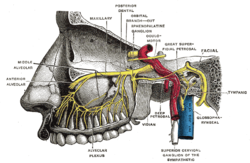
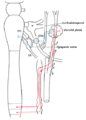
The otic ganglion is a small parasympathetic ganglion located immediately below the foramen ovale in the infratemporal fossa and on the medial surface of the mandibular nerve. It is functionally associated with the glossopharyngeal nerve and innervates the parotid gland for salivation.

The auriculotemporal nerve is a sensory branch of the mandibular nerve (CN V3) that runs with the superficial temporal artery and vein, and provides sensory innervation to parts of the external ear, scalp, and temporomandibular joint. The nerve also conveys post-ganglionic parasympathetic fibres from the otic ganglion to the parotid gland.

Chorda tympani is a branch of the facial nerve that carries gustatory (taste) sensory innervation from the front of the tongue and parasympathetic (secretomotor) innervation to the submandibular and sublingual salivary glands.

The tympanic cavity is a small cavity surrounding the bones of the middle ear. Within it sit the ossicles, three small bones that transmit vibrations used in the detection of sound.

The greater petrosal nerve is a nerve of the head mainly containing pre-ganglionic parasympathetic fibres which ultimately synapse in the pterygopalatine ganglion. It branches from the facial nerve and is derived from the parasympathetic part of the nervus intermedius component of CN VII, with its cell bodies located in the superior salivary nucleus. In the connective tissue substance of the foramen lacerum, the greater petrosal nerve unites with the (sympathetic) deep petrosal nerve to form the nerve of the pterygoid canal which proceeds to the pterygopalatine ganglion.
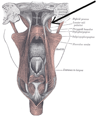
The tensor veli palatini muscle is a thin, triangular muscle of the head that tenses the soft palate and opens the Eustachian tube to equalise pressure in the middle ear.
Petrosal nerve may refer to:

The petrous part of the temporal bone is pyramid-shaped and is wedged in at the base of the skull between the sphenoid and occipital bones. Directed medially, forward, and a little upward, it presents a base, an apex, three surfaces, and three angles, and houses in its interior the components of the inner ear. The petrous portion is among the most basal elements of the skull and forms part of the endocranium. Petrous comes from the Latin word petrosus, meaning "stone-like, hard". It is one of the densest bones in the body. In other mammals, it is a separate bone, the petrosal bone.
The lesser petrosal nerve is the general visceral efferent (GVE) nerve conveying pre-ganglionic parasympathetic secretomotor fibers for the parotid gland from the tympanic plexus to the otic ganglion. It passes out of the tympanic cavity through the petrous part of the temporal bone into the middle cranial fossa of the cranial cavity, then exits the cranial cavity through its own canaliculus to reach the infratemporal fossa.
The superior ganglion of the glossopharyngeal nerve is a sensory ganglion of the peripheral nervous system. It is located within the jugular foramen where the glossopharyngeal nerve exits the skull. It is smaller than and superior to the inferior ganglion of the glossopharyngeal nerve.

Parasympathetic ganglia are the autonomic ganglia of the parasympathetic nervous system. Most are small terminal ganglia or intramural ganglia, so named because they lie near or within (respectively) the organs they innervate. The exceptions are the four paired parasympathetic ganglia of the head and neck.

The tympanic plexus is a nerve plexus within the tympanic cavity formed upon the promontory of tympanic cavity by the tympanic nerve, and the superior and inferior caroticotympanic nerves.
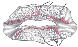
The labial glands are minor salivary glands situated between the mucous membrane and the orbicularis oris around the orifice of the mouth.

The salivatory nuclei are pre-ganglionic parasympathetic neurons in the caudal pons representing the general visceral efferent (GVE) cranial nerve nuclei giving rise to axons which join the facial nerve and glossopharyngeal nerve to reach and innervate the salivary as well as lacrimal glands. The nuclei may also be involved in parasympathetic control of head vasculature.

The following outline is provided as an overview of and topical guide to human anatomy:

