The middle ear is the portion of the ear medial to the eardrum, and distal to the oval window of the cochlea.

The oval window is a membrane-covered opening from the middle ear to the cochlea of the inner ear.
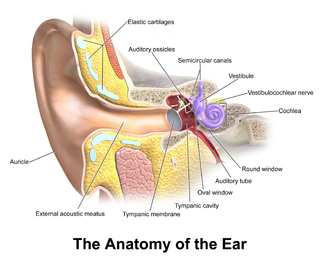
In the anatomy of humans and various other tetrapods, the eardrum, also called the tympanic membrane or myringa, is a thin, cone-shaped membrane that separates the external ear from the middle ear. Its function is to transmit sound from the air to the ossicles inside the middle ear, and then to the oval window in the fluid-filled cochlea. Hence, it ultimately converts and amplifies vibration in the air to vibration in cochlear fluid. The malleus bone bridges the gap between the eardrum and the other ossicles.

The temporal bones are situated at the sides and base of the skull, and lateral to the temporal lobes of the cerebral cortex.

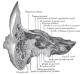
The tympanic cavity is a small cavity surrounding the bones of the middle ear. Within it sit the ossicles, three small bones that transmit vibrations used in the detection of sound.
The tensor veli palatini muscle is a thin, triangular muscle of the head that tenses the soft palate and opens the Eustachian tube to equalise pressure in the middle ear.
The round window is one of the two openings from the middle ear into the inner ear. It is sealed by the secondary tympanic membrane, which vibrates with opposite phase to vibrations entering the inner ear through the oval window. It allows fluid in the cochlea to move, which in turn ensures that hair cells of the basilar membrane will be stimulated and that audition will occur.

The anterior ethmoidal artery is a branch of the ophthalmic artery in the orbit. It exits the orbit through the anterior ethmoidal foramen alongside the anterior ethmoidal nerve. It contributes blood supply to the ethmoid sinuses, frontal sinuses, the dura mater, lateral nasal wall, and nasal septum. It issues a meningeal branch, and nasal branches.

The mastoid part of the temporal bone is the posterior (back) part of the temporal bone, one of the bones of the skull. Its rough surface gives attachment to various muscles and it has openings for blood vessels. From its borders, the mastoid part articulates with two other bones.

The tympanic part of the temporal bone is a curved plate of bone lying below the squamous part of the temporal bone, in front of the mastoid process, and surrounding the external part of the ear canal.

The facial canal is a Z-shaped canal in the temporal bone of the skull. It extends between the internal acoustic meatus and stylomastoid foramen. It transmits the facial nerve.

The tympanic nerve is a branch of the glossopharyngeal nerve found near the ear. It gives sensation to the middle ear, the Eustachian tube, the parotid gland, and mastoid air cells. It gives parasympathetic to supply to the parotid gland via the otic ganglion and the auriculotemporal nerve.

The pyramidal eminence is a hollow conical projection upon the posterior wall of the tympanic cavity of the middle ear. The stapedius muscle arises in the hollow of the eminence and its tendon exits through its apex.
The aditus to mastoid antrum, is a large, irregular opening upon the posterior wall of the tympanic cavity by which the mastoid antrum communicates with the epitympanic recess of the tympanic cavity. The walls of the antrum are lined by mucosa which is continuous with that lining the mastoid cells and tympanic cavity.

The tympanic plexus is a nerve plexus within the tympanic cavity formed upon the promontory of tympanic cavity by the tympanic nerve, and the superior and inferior caroticotympanic nerves.

The vestibule is the central part of the bony labyrinth in the inner ear, and is situated medial to the eardrum, behind the cochlea, and in front of the three semicircular canals.
The caroticotympanic nerves are post-ganglionic sympathetic branches from the internal carotid plexus which leave the carotid canal through the wall of this canal to enter the tympanic cavity and participate in the formation of the tympanic plexus upon the promontory of tympanic cavity. They travel with the caroticotympanic artery.

The following outline is provided as an overview of and topical guide to human anatomy:

The inferior tympanic artery is a small branch of the ascending pharyngeal artery which passes through the tympanic canaliculus alongside the tympanic branch of glossopharyngeal nerve to reach and provide arterial supply to the medial wall of the tympanic cavity where it forms anastomoses with the other tympanic arteries.

Many anatomical terms descriptive of bone are defined in anatomical terminology, and are often derived from Greek and Latin. Bone in the human body is categorized into long bone, short bone, flat bone, irregular bone and sesamoid bone.