
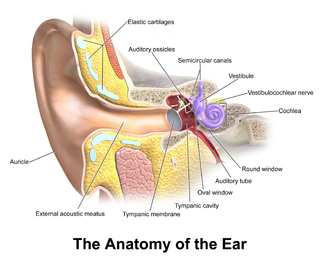
The middle ear is the portion of the ear medial to the eardrum, and distal to the oval window of the cochlea.
The outer ear, external ear, or auris externa is the external part of the ear, which consists of the auricle and the ear canal. It gathers sound energy and focuses it on the eardrum.
The ossicles are three bones in either middle ear that are among the smallest bones in the human body. They serve to transmit sound vibrations sent from the ear drum to the fluid-filled labyrinth (cochlea). The absence of the auditory ossicles would constitute a moderate-to-severe hearing loss. The term "ossicle" literally means "tiny bone". Though the term may refer to any small bone throughout the body, it typically refers to the malleus, incus, and stapes of the middle ear.
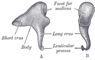
The incus or anvil in the ear is one of three small bones (ossicles) in the middle ear. The incus receives vibrations from the malleus, to which it is connected laterally, and transmits these to the stapes medially. The incus is named for its resemblance to an anvil.
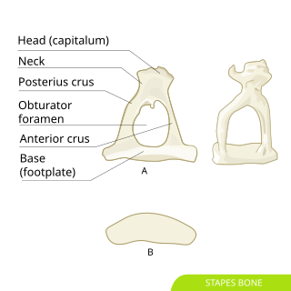
The stapes or stirrup is a bone in the middle ear of humans and other tetrapods which is involved in the conduction of sound vibrations to the inner ear. This bone is connected to the oval window by its annular ligament, which allows the footplate to transmit sound energy through the oval window into the inner ear. The stapes is the smallest and lightest bone in the human body, and is so-called because of its resemblance to a stirrup.
In the anatomy of humans and various other tetrapods, the eardrum, also called the tympanic membrane or myringa, is a thin, cone-shaped membrane that separates the external ear from the middle ear. Its function is to transmit changes in pressure of sound from the air to the ossicles inside the middle ear, and thence to the oval window in the fluid-filled cochlea. The ear thereby converts and amplifies vibration in the air to vibration in cochlear fluid. The malleus bone bridges the gap between the eardrum and the other ossicles.

The fibula or calf bone is a leg bone on the lateral side of the tibia, to which it is connected above and below. It is the smaller of the two bones and, in proportion to its length, the most slender of all the long bones. Its upper extremity is small, placed toward the back of the head of the tibia, below the knee joint and excluded from the formation of this joint. Its lower extremity inclines a little forward, so as to be on a plane anterior to that of the upper end; it projects below the tibia and forms the lateral part of the ankle joint.

The quadrate bone is a skull bone in most tetrapods, including amphibians, sauropsids, and early synapsids.

An ear is the organ that enables hearing and body balance using the vestibular system. In mammals, the ear is usually described as having three parts: the outer ear, the middle ear and the inner ear. The outer ear consists of the pinna and the ear canal. Since the outer ear is the only visible portion of the ear in most animals, the word "ear" often refers to the external part alone. The middle ear includes the tympanic cavity and the three ossicles. The inner ear sits in the bony labyrinth, and contains structures which are key to several senses: the semicircular canals, which enable balance and eye tracking when moving; the utricle and saccule, which enable balance when stationary; and the cochlea, which enables hearing. The ear canal is cleaned via earwax, which naturally migrates to the auricle. The ears of vertebrates are placed somewhat symmetrically on either side of the head, an arrangement that aids sound localization.

The tympanic cavity is a small cavity surrounding the bones of the middle ear. Within it sit the ossicles, three small bones that transmit vibrations used in the detection of sound.

The pharyngeal arches, also known as visceral arches, are transient structures seen in the embryonic development of humans and other vertebrates, that are recognisable precursors for many structures. In fish, the arches support the gills and are known as the branchial arches, or gill arches.

The oblique popliteal ligament is a broad, flat, fibrous ligament on the posterior knee. It is an extension of the tendon of the semimembranosus muscle. It attaches onto the intercondylar fossa and lateral condyle of the femur. It reinforces the posterior central portion of the knee joint capsule.
The superior ligament of the incus is a fibrous band that crosses from the body of the incus to the roof of the tympanic cavity just posterior to the superior ligament of the malleus.

The evolution of mammalian auditory ossicles was an evolutionary process that resulted in the formation of the mammalian middle ear, where the three middle ear bones or ossicles, namely the incus, malleus and stapes, are a defining characteristic of mammals. The event is well-documented and important academically as a demonstration of transitional forms and exaptation, the re-purposing of existing structures during evolution.

The epitympanic recess is the portion of the tympanic cavity situated superior to the tympanic membrane. The recess lodges the head of malleus, and the body of incus.

The following outline is provided as an overview of and topical guide to human anatomy:

Hearing, or auditory perception, is the ability to perceive sounds through an organ, such as an ear, by detecting vibrations as periodic changes in the pressure of a surrounding medium. The academic field concerned with hearing is auditory science.

The incudomalleolar joint or articulatio incudomallearis is a small synovial joint between the malleus (hammer) and the incus (anvil). The joint's function is to transfer vibrations between the ossicles in the middle ear, which is perceived as sound. Contrary to other synovial joints the movement is very limited. All of the ossicles move more or less as a unit, at least at low frequencies.
Ligament of incus may refer to:

The malleus, or hammer, is a hammer-shaped small bone or ossicle of the middle ear. It connects with the incus, and is attached to the inner surface of the eardrum. The word is Latin for 'hammer' or 'mallet'. It transmits the sound vibrations from the eardrum to the incus (anvil).