
The inner ear is the innermost part of the vertebrate ear. In vertebrates, the inner ear is mainly responsible for sound detection and balance. In mammals, it consists of the bony labyrinth, a hollow cavity in the temporal bone of the skull with a system of passages comprising two main functional parts:

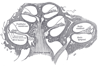
The cochlea is the part of the inner ear involved in hearing. It is a spiral-shaped cavity in the bony labyrinth, in humans making 2.75 turns around its axis, the modiolus. A core component of the cochlea is the organ of Corti, the sensory organ of hearing, which is distributed along the partition separating the fluid chambers in the coiled tapered tube of the cochlea.

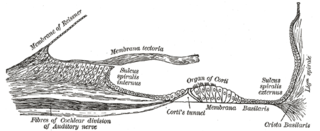
The basilar membrane is a stiff structural element within the cochlea of the inner ear which separates two liquid-filled tubes that run along the coil of the cochlea, the scala media and the scala tympani. The basilar membrane moves up and down in response to incoming sound waves, which are converted to traveling waves on the basilar membrane.
The organ of Corti, or spiral organ, is the receptor organ for hearing and is located in the mammalian cochlea. This highly varied strip of epithelial cells allows for transduction of auditory signals into nerve impulses' action potential. Transduction occurs through vibrations of structures in the inner ear causing displacement of cochlear fluid and movement of hair cells at the organ of Corti to produce electrochemical signals.

The auditory system is the sensory system for the sense of hearing. It includes both the sensory organs and the auditory parts of the sensory system.

An ear is the organ that enables hearing and body balance using the vestibular system. In mammals, the ear is usually described as having three parts: the outer ear, the middle ear and the inner ear. The outer ear consists of the pinna and the ear canal. Since the outer ear is the only visible portion of the ear in most animals, the word "ear" often refers to the external part alone. The middle ear includes the tympanic cavity and the three ossicles. The inner ear sits in the bony labyrinth, and contains structures which are key to several senses: the semicircular canals, which enable balance and eye tracking when moving; the utricle and saccule, which enable balance when stationary; and the cochlea, which enables hearing. The ear is a self cleaning organ through its relationship with earwax and the ear canals. The ears of vertebrates are placed somewhat symmetrically on either side of the head, an arrangement that aids sound localization.
Endolymph is the fluid contained in the membranous labyrinth of the inner ear. The major cation in endolymph is potassium, with the values of sodium and potassium concentration in the endolymph being 0.91 mM and 154 mM, respectively. It is also called Scarpa's fluid, after Antonio Scarpa.
Presbycusis, or age-related hearing loss, is the cumulative effect of aging on hearing. It is a progressive and irreversible bilateral symmetrical age-related sensorineural hearing loss resulting from degeneration of the cochlea or associated structures of the inner ear or auditory nerves. The hearing loss is most marked at higher frequencies. Hearing loss that accumulates with age but is caused by factors other than normal aging is not presbycusis, although differentiating the individual effects of distinct causes of hearing loss can be difficult.
Perilymph is an extracellular fluid located within the inner ear. It is found within the scala tympani and scala vestibuli of the cochlea. The ionic composition of perilymph is comparable to that of plasma and cerebrospinal fluid. The major cation in perilymph is sodium, with the values of sodium and potassium concentration in the perilymph being 138 mM and 6.9 mM, respectively. It is also named Cotunnius' liquid and liquor cotunnii for Domenico Cotugno.
The cochlear nerve is one of two parts of the vestibulocochlear nerve, a cranial nerve present in amniotes, the other part being the vestibular nerve. The cochlear nerve carries auditory sensory information from the cochlea of the inner ear directly to the brain. The other portion of the vestibulocochlear nerve is the vestibular nerve, which carries spatial orientation information to the brain from the semicircular canals, also known as semicircular ducts.
The cochlear duct is an endolymph filled cavity inside the cochlea, located between the tympanic duct and the vestibular duct, separated by the basilar membrane and the vestibular membrane respectively. The cochlear duct houses the organ of Corti.
The vestibular membrane, vestibular wall or Reissner's membrane is a membrane inside the cochlea of the inner ear. It separates the cochlear duct from the vestibular duct. It helps to transmit vibrations from fluid in the vestibular duct to the cochlear duct. Together with the basilar membrane, it creates a compartment in the cochlea filled with endolymph, which is important for the function of the spiral organ of Corti. It allows nutrients to travel from the perilymph to the endolymph of the membranous labyrinth. It may be damaged in Ménière's disease. It is named after the German anatomist Ernst Reissner.

A kinocilium is a special type of cilium on the apex of hair cells located in the sensory epithelium of the vertebrate inner ear. Contrasting with stereocilia, which are numerous, there is only one kinocilium on each hair cell. The kinocilium can be identified by its apical position as well as its enlarged tip.

The crista ampullaris is the sensory organ of rotation. They are found in the ampullae of each of the semicircular canals of the inner ear, meaning that there are three pairs in total. The function of the crista ampullaris is to sense angular acceleration and deceleration.

The vestibule is the central part of the bony labyrinth in the inner ear, and is situated medial to the eardrum, behind the cochlea, and in front of the three semicircular canals.
The spiral ligament is a fibrous cushion located between the stria vascularis and the bony otic capsule.
The neural encoding of sound is the representation of auditory sensation and perception in the nervous system. The complexities of contemporary neuroscience are continually redefined. Thus what is known of the auditory system has been continually changing. The encoding of sounds includes the transduction of sound waves into electrical impulses along auditory nerve fibers, and further processing in the brain.
Claudius cells are considered as supporting cells within the organ of Corti in the cochlea. These cells extend from Hensen's cells to the spiral prominence epithelium, forming the outer sulcus. They are in direct contact with the endolymph of the cochlear duct. These cells are sealed via tight junctions that prevent flow of endolymph between them. Boettcher cells are located immediately under Claudius cells in the lower turn of the cochlea.
The reticular membrane is a thin, stiff lamina that extends from the outer hair cells to the Hensen's cells. The RM is composed of "minute-fiddle-shaped cuticular structures" called the phalangeal extensions of the outer hair cells, interspaced with extensions coming from the outer phalangeal cells. The RM separates endolymph in the cochlear duct from underlying corticolymph and perilymph of the scala tympani.
Dark cells are specialized nonsensory epithelial cells found on either side of the vestibular organs and lining the endolymphatic space. These dark-cell areas in the vestibular organ are structures involved in the production of endolymph, an inner ear fluid, secreting potassium towards the endolymphatic fluid. Dark cells take part in fluid homeostasis to preserve the unique high-potassium and low-sodium content of the endolymph and also maintain the calcium homeostasis of the inner ear.
