Saccule

Last updated
Saccule
Blausen 0329 EarAnatomy InternalEar.png
Inner ear, showing saccule near center.
Bigotolith.jpg
illustration of otolith organs showing detail of utricle, otoconia, endolymph, cupula, Macula of saccule, hair cell filaments, and saccular nerve
Details
Part of Inner ear
System Balance
Identifiers
Latin sacculus
TA98 A15.3.03.065
TA2 7001
FMA 61116
Anatomical terminology

The saccule is a bed of sensory cells in the inner ear. It translates head movements into neural impulses for the brain to interpret. The saccule detects linear accelerations and head tilts in the vertical plane. When the head moves vertically, the sensory cells of the saccule are disturbed and the neurons connected to them begin transmitting impulses to the brain. These impulses travel along the vestibular portion of the eighth cranial nerve to the vestibular nuclei in the brainstem.

Contents

The vestibular system is important in maintaining balance, or equilibrium. The vestibular system includes the saccule, utricle, and the three semicircular canals. The vestibule is the name of the fluid-filled, membranous duct that contains these organs of balance. The vestibule is encased in the temporal bone of the skull.

Structure

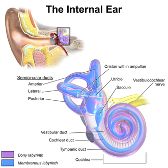
The saccule, or sacculus, is the smaller of the two vestibular sacs. It is globular in form and lies in the recessus sphæricus near the opening of the vestibular duct of the cochlea. Its cavity does not directly communicate with that of the utricle. The anterior part of the saccule exhibits an oval thickening, the macula acustica sacculi, or macula, to which are distributed the saccular filaments of the vestibular branch of the vestibulocochlear nerve, also known as the statoacoustic nerve or cranial nerve VIII.

Within the macula are hair cells, each having a hair bundle on the apical aspect. The hair bundle is composed of a single kinocilium and many (at least 70) stereocilia. Stereocilia are connected to mechanically gated ion channels in the hair cell plasma membrane via tip links. Supporting cells interdigitate between hair cells and secrete the otolithic membrane, a thick, gelatinous layer of glycoprotein. Covering the surface of the otolithic membrane are otoliths, which are crystals of calcium carbonate. For this reason, the saccule is sometimes called an "otolithic organ."

From the posterior wall of the saccule is given off a canal, the ductus endolymphaticus (endolymphatic duct). This duct is joined by the ductus utriculosaccularis, and then passes along the aquæductus vestibuli and ends in a blind pouch saccus endolymphaticus (endolymphatic sac) on the posterior surface of the petrous portion of the temporal bone, where it is in contact with the dura mater.

From the lower part of the saccule a short tube, the canalis reuniens of Hensen, passes downward and opens into the ductus cochlearis near its vestibular extremity.

Both the utricle and the saccule provide information about acceleration. The difference between them is that the utricle is more sensitive to horizontal acceleration, whereas the saccule is more sensitive to vertical acceleration.

Function

Saccule
Components of the inner ear

The saccule gathers sensory information to orient the body in space. It primarily gathers information about linear movement in the vertical plane, including the force due to gravity. The saccule, like the utricle, provides information to the brain about head position when it is not moving. [1] The structures that enable the saccule to gather this vestibular information are the hair cells. The 2 by 3 mm patch of hair cells and supporting cells are called a macula. Each hair cell of a macula has 40 to 70 stereocilia and one true cilium called a kinocilium. The stereocilia are oriented by the striola, a curved ridge that runs through the middle of the macula; in the saccule they are oriented away from the striola [2] The tips of the stereocilia and kinocilium are embedded in a gelatinous otolithic membrane. This membrane is weighted with protein-calcium carbonate granules called otoliths, which add to the weight and inertia of the membrane and enhance the sense of gravity and motion. [3]

Not much is known of how this organ is used in other species. Research has shown, like songbirds, females in some species of fish show seasonal variation in auditory processing and the sensitivity of the saccule of females peaks during the breeding season. This is due to an increase in the density of saccular hair cells, partly resulting from reduced apoptosis. [4] The increase the hair cells make also increase the sensitivity to male mating calls. An example of this is seen in Porichthys notatus, or plainfin midshipman fish.

Clinical significance

Assessment

Saccular function can be assessed by the cervical vestibular evoked myogenic potential (cVEMP). This is a middle latency (P1 between 12-20 ms) waveform denoting inhibition of the sternocleidomastoid (SCM) muscle ipsilateral to the stimulus. While not truly a unilateral reflex (response waveforms can be detected in the SCM contralateral to the stimulus in approximately 40% of cases), cVEMPs are more unilateral than the closely related ocular vestibular evoked myogenic potential (oVEMP). The most reliable points on the cVEMP waveform are known as P1 and N1. Of all waveform characteristics, P1-N1 amplitude is the most reliable and clinically relevant. cVEMP amplitude is linearly dependent upon stimulus intensity and is most reliably elicited with a loud (generally at or above 95 dB nHL) click or tone burst. The cVEMP can also be said to be low-frequency tuned, with largest amplitudes in response to 500–750 Hz tonebursts. This myogenic potential is felt to assess saccular function, because the response is present in completely deafened ears and because it is routed through the inferior vestibular nerve, which is known to dominantly innervate the saccule. . [5]

Role in evolution of the ear

Research suggests that in vertebrate evolution, sensory cells became specialized as gravistatic sensors after they became assembled to form the ear. After this aggregation, growth, including duplication and segregation of existing neurosensory epithelia, gave rise to new epithelia and can be appreciated by comparing sensory epithelia from the inner ears of different vertebrates and their innervation by different neuronal populations. Novel directions of differentiation were apparently further expanded by incorporating unique molecular modules in newly developed sensory epithelia. For example, the saccule gave rise to the auditory epithelium and corresponding neuronal population of tetrapods, starting possibly in an aquatic environment. [6]

See also

Related Research Articles

<span class="mw-page-title-main">Inner ear</span> Innermost part of the vertebrate ear

The inner ear is the innermost part of the vertebrate ear. In vertebrates, the inner ear is mainly responsible for sound detection and balance. In mammals, it consists of the bony labyrinth, a hollow cavity in the temporal bone of the skull with a system of passages comprising two main functional parts:

<span class="mw-page-title-main">Sense of balance</span> Physiological sense regarding posture

The sense of balance or equilibrioception is the perception of balance and spatial orientation. It helps prevent humans and nonhuman animals from falling over when standing or moving. Equilibrioception is the result of a number of sensory systems working together; the eyes, the inner ears, and the body's sense of where it is in space (proprioception) ideally need to be intact.

<span class="mw-page-title-main">Cochlea</span> Snail-shaped part of inner ear involved in hearing

The cochlea is the part of the inner ear involved in hearing. It is a spiral-shaped cavity in the bony labyrinth, in humans making 2.75 turns around its axis, the modiolus. A core component of the cochlea is the organ of Corti, the sensory organ of hearing, which is distributed along the partition separating the fluid chambers in the coiled tapered tube of the cochlea.

<span class="mw-page-title-main">Vestibulocochlear nerve</span> Cranial nerve VIII, for hearing and balance

The vestibulocochlear nerve or auditory vestibular nerve, also known as the eighth cranial nerve, cranial nerve VIII, or simply CN VIII, is a cranial nerve that transmits sound and equilibrium (balance) information from the inner ear to the brain. Through olivocochlear fibers, it also transmits motor and modulatory information from the superior olivary complex in the brainstem to the cochlea.

<span class="mw-page-title-main">Semicircular canals</span> Organ located in innermost part of ear

The semicircular canals are three semicircular interconnected tubes located in the innermost part of each ear, the inner ear. The three canals are the lateral, anterior and posterior semicircular canals. They are the part of the bony labyrinth, a periosteum-lined cavity on the petrous part of the temporal bone filled with perilymph.

<span class="mw-page-title-main">Utricle (ear)</span> Membranous labyrinth in the vestibule of ear

The utricle and saccule are the two otolith organs in the vertebrate inner ear. They are part of the balancing system in the vestibule of the bony labyrinth. They use small stones and a viscous fluid to stimulate hair cells to detect motion and orientation. The utricle detects linear accelerations and head-tilts in the horizontal plane. The word utricle comes from Latin uter 'leather bag'.

<span class="mw-page-title-main">Vestibular system</span> Sensory system that facilitates body balance

The vestibular system, in vertebrates, is a sensory system that creates the sense of balance and spatial orientation for the purpose of coordinating movement with balance. Together with the cochlea, a part of the auditory system, it constitutes the labyrinth of the inner ear in most mammals.

<span class="mw-page-title-main">Otolith</span> Inner-ear structure in vertebrates which detects acceleration

An otolith, also called statoconium or otoconium or statolith, is a calcium carbonate structure in the saccule or utricle of the inner ear, specifically in the vestibular system of vertebrates. The saccule and utricle, in turn, together make the otolith organs. These organs are what allows an organism, including humans, to perceive linear acceleration, both horizontally and vertically (gravity). They have been identified in both extinct and extant vertebrates.

<span class="mw-page-title-main">Stereocilia (inner ear)</span> Mechanosensing organelles of hair cells

In the inner ear, stereocilia are the mechanosensing organelles of hair cells, which respond to fluid motion in numerous types of animals for various functions, including hearing and balance. They are about 10–50 micrometers in length and share some similar features of microvilli. The hair cells turn the fluid pressure and other mechanical stimuli into electric stimuli via the many microvilli that make up stereocilia rods. Stereocilia exist in the auditory and vestibular systems.

The vestibular ganglion is a collection of cell bodies belonging to first order sensory neurons of the vestibular nerve. It is located within the internal auditory canal.

<span class="mw-page-title-main">Ampullary cupula</span>

The ampullary cupula, or cupula, is a structure in the vestibular system, providing the sense of spatial orientation.

<span class="mw-page-title-main">Otolithic membrane</span>

The otolithic membrane is a fibrous structure located in the vestibular system of the inner ear. It plays a critical role in the brain's interpretation of equilibrium. The membrane serves to determine if the body or the head is tilted, in addition to the linear acceleration of the body. The linear acceleration could be in the horizontal direction as in a moving car or vertical acceleration such as that felt when an elevator moves up or down.

<span class="mw-page-title-main">Kinocilium</span> Sensory hair in the inner ear

A kinocilium is a special type of cilium on the apex of hair cells located in the sensory epithelium of the vertebrate inner ear. Contrasting with stereocilia, which are numerous, there is only one kinocilium on each hair cell. The kinocilium can be identified by its apical position as well as its enlarged tip.

<span class="mw-page-title-main">Crista ampullaris</span> Sensory organ in the inner ear

The crista ampullaris is the sensory organ of rotation. They are found in the ampullae of each of the semicircular canals of the inner ear, meaning that there are three pairs in total. The function of the crista ampullaris is to sense angular acceleration and deceleration.

<span class="mw-page-title-main">Otic vesicle</span> Two sac-like invaginations formed and subsequently closed off during embryonic development

Otic vesicle, or auditory vesicle, consists of either of the two sac-like invaginations formed and subsequently closed off during embryonic development. It is part of the neural ectoderm, which will develop into the membranous labyrinth of the inner ear. This labyrinth is a continuous epithelium, giving rise to the vestibular system and auditory components of the inner ear. During the earlier stages of embryogenesis, the otic placode invaginates to produce the otic cup. Thereafter, the otic cup closes off, creating the otic vesicle. Once formed, the otic vesicle will reside next to the neural tube medially, and on the lateral side will be paraxial mesoderm. Neural crest cells will migrate rostral and caudal to the placode.

The saccule is the smaller sized vestibular sac ; it is globular in form, and lies in the recessus sphæricus near the opening of the scala vestibuli of the cochlea. Its anterior part exhibits an oval thickening, the macula of saccule, to which are distributed the saccular filaments of the acoustic nerve.

<span class="mw-page-title-main">Arthur Böttcher</span> Baltic German scientist

Jakob Ernst Arthur Böttcher was a Baltic German pathologist and anatomist who was a native of Bauska, in what was then the Courland Governorate. He worked primarily within the Russian Empire.

The vestibular evoked myogenic potential is a neurophysiological assessment technique used to determine the function of the otolithic organs of the inner ear. It complements the information provided by caloric testing and other forms of inner ear testing. There are two different types of VEMPs. One is the oVEMP and another is the cVEMP. The oVEMP measures integrity of the utricule and superior vestibular nerve and the cVemp measures the saccule and the inferior vestibular nerve.

Electrocochleography is a technique of recording electrical potentials generated in the inner ear and auditory nerve in response to sound stimulation, using an electrode placed in the ear canal or tympanic membrane. The test is performed by an otologist or audiologist with specialized training, and is used for detection of elevated inner ear pressure or for the testing and monitoring of inner ear and auditory nerve function during surgery.

References

  1. How Our Balance System Works American Speech-Language-Hearing Association, 2013
  2. Fitzakerly, Janet University of Minnesota Medical School Deluth, February 10, 2013
  3. Saladin, Kenneth S. Anatomy & Physiology The Unity of Form and Function. 6th Ed. New York: McGraw Hill, 2012. 605-609. Print.
  4. Coflfin B. Allison Saccular-Specific Hair Cell Addition Correlates with Reproductive State-Dependent Changes in the Auditory Saccular Sensitivity of a Vocal Fish Journal of Neuroscience, January 25, 2012
  5. Cushing,& Lynn, S. (2008). "Relationship between sensorineural hearing loss and vestibular and balance function in children." (Master's thesis, University of Toronto, Canada)Retrieved from url:
  6. Duncan, Jeremy Shane (2012). Cochlear neurosensory specification and competence: you gata have Gata (PhD thesis). University of Iowa.