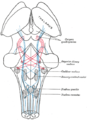
Lateral lemniscus

Last updated
Lateral lemniscus
Lateral lemniscus.PNG
Lateral lemniscus in red, as it connects the cochlear nucleus, superior olivary nucleus and the inferior colliculus. Seen from behind.
Details
Identifiers
Latin lemniscus lateralis
NeuroNames 609
NeuroLex ID birnlex_976
TA98 A14.1.05.317
A14.1.08.670
A14.1.06.204
TA2 5866
FMA 72502
Anatomical terms of neuroanatomy

The lateral lemniscus is a tract of axons in the brainstem that carries information about sound from the cochlear nucleus to various brainstem nuclei and ultimately the contralateral inferior colliculus of the midbrain. Three distinct, primarily inhibitory, cellular groups are located interspersed within these fibers, and are thus named the nuclei of the lateral lemniscus.

Contents

Connections

There are three small nuclei on each of the lateral lemnisci:

Fibers leaving these brainstem nuclei ascending to the inferior colliculus rejoin the lateral lemniscus. In that sense, this is not a 'lemniscus' in the true sense of the word (second order, decussated sensory axons), as there is third (and out of the lateral superior olive, fourth) order information coming out of some of these brainstem nuclei.

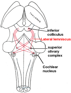
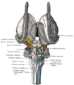
The lateral lemniscus is located where the cochlear nuclei and the pontine reticular formation (PRF) crossover. The PRF descends the reticulospinal tract where it innervates motor neurons and spinal interneurons. It is the main auditory tract in the brainstem that connects the superior olivary complex (SOC) with the inferior colliculus (IC). The dorsal cochlear nucleus (DCN) has input from the LL and output to the contralateral LL via the ipsilateral and contralateral Dorsal Acoustic Stria.

The two lemnisci communicate via the commissural fibers of Probst.

Nuclei of the lateral lemniscus

The function of the complex of Nuclei of the lateral lemniscus is not known; however it has good temporal resolution compared to other cells higher than the cochlear nuclei and is sensitive to both timing and amplitude changes in sound. It is also involved in the acoustic startle reflex; the most likely region for this being the VNLL.

DNLL

The cells of the DNLL respond best to bilateral inputs, and have onset and complexity tuned sustained responses. The nucleus is primarily GABAergic, [1] and projects bilaterally to the inferior colliculus, and contralaterally to the DNLL, with different populations of cells projecting to each IC. [2]

In rat, the DNLL has a prominent columnar organization. Nearly all neurons are stained for GABA, especially in the central part of the nucleus, and the remaining GABA negative cells are interspersed with the positive, and often stain for glycine. Two populations of GABA+ cells are visible: larger, lightly stained cells that project to the contralateral IC, and smaller, darker stained cells that project ipsilaterally. GABAergic axon terminals form dense groups surrounded by GABA-lemniscal fibers throughout the nucleus, and synapse on both somata and in the neuropil. Glycinergic axon terminals, on the other hand, are more finely localized, with the majority of recipient neurons located laterally in the nucleus. [3]

INLL

INLL also has little spontaneous activity and broad tuning curves. The temporal responses are significantly different from cells of the VNLL.

This structure is greatly hypertrophied in the rat, forming a prominent bulge on the surface of the brainstem. GAD, GABA, and Glycine staining reveals several distinct regions that are not evident in standard cytoarchitectural preparations. A modest number of GABA-stained neurons are arranged in small groups, generally in the center of the nucleus, whereas glycine-stained neurons are more common and widely dispersed, with regional concentrations in the dorsolateral and ventrolateral portions of the nucleus. Most GABA+ cells are gly+ as well. [1] [broken footnote]

VNLL

Sound in the contralateral ear leads to the strongest responses in the VNLL, which deals with some temporary processing. The VNLL may also be essential to the IC’s decoding of amplitude modulated sounds.

VNLL cells have little spontaneous activity, broad and moderately complex tuning curves; they have both phasic and tonic responses and are involved in temporal processing.

In rat, the VNLL is composed of two subdivisions, the ventral (columnar) and dorsal (non columnar) regions. The columnar region contains many glycine-positive (0 GABA+) neurons, whereas the dorsal region contains clusters of GABA+ neurons intermingled with gly+ cells, with some cells containing both. [1]

Inputs and outputs to nuclei

The table below shows that each of the nuclei have a complicated arrangement of ipsilateral and contralateral afferent inputs and outputs:[ citation needed ]

NucleusInputOutput
 ContralateralIpsilateralContralateralIpsilateral
VNLLAnterior and posterior ventral cochlear nucleiMedial nucleus of the trapezoid body Inferior Colliculus
DNLL
INLLAnterior and posterior Ventral Cochlear NucleusMedial nucleus of the trapezoid body Medial Geniculate body
Inferior Colliculus
DNLLAnterior Ventral
Cochlear nucleus (and Bilateral)
Medial superior Olivary Nucleus
Lateral Superior Olivary Nucleus (and Bilateral)
DNLL
Inferior Colliculus
Mid brain reticular formation
Superior Olivary Complex
Inferior Colliculus
 Medial Geniculate Body
Mid brain reticular formation
Superior Olivary Complex

Additional images

Related Research Articles

<span class="mw-page-title-main">Medulla oblongata</span> Structure of the brain stem

The medulla oblongata or simply medulla is a long stem-like structure which makes up the lower part of the brainstem. It is anterior and partially inferior to the cerebellum. It is a cone-shaped neuronal mass responsible for autonomic (involuntary) functions, ranging from vomiting to sneezing. The medulla contains the cardiac, respiratory, vomiting and vasomotor centers, and therefore deals with the autonomic functions of breathing, heart rate and blood pressure as well as the sleep–wake cycle.

Articles related to anatomy include:

<span class="mw-page-title-main">Brainstem</span> Posterior part of the brain, adjoining and structurally continuous

The brainstem is the stalk-like part of the brain that interconnects the cerebrum and diencephalon with the spinal cord. In the human brain, the brainstem is composed of the midbrain, the pons, and the medulla oblongata. The midbrain is continuous with the thalamus of the diencephalon through the tentorial notch.

<span class="mw-page-title-main">Lateral geniculate nucleus</span> Component of the visual system in the brains thalamus

In neuroanatomy, the lateral geniculate nucleus is a structure in the thalamus and a key component of the mammalian visual pathway. It is a small, ovoid, ventral projection of the thalamus where the thalamus connects with the optic nerve. There are two LGNs, one on the left and another on the right side of the thalamus. In humans, both LGNs have six layers of neurons alternating with optic fibers.

<span class="mw-page-title-main">Spinothalamic tract</span> Sensory pathway from the skin to the thalamus

The spinothalamic tract is a part of the anterolateral system or the ventrolateral system, a sensory pathway to the thalamus. From the ventral posterolateral nucleus in the thalamus, sensory information is relayed upward to the somatosensory cortex of the postcentral gyrus.

<span class="mw-page-title-main">Dorsal column–medial lemniscus pathway</span> Sensory spinal pathway

The dorsal column–medial lemniscus pathway (DCML) is a sensory pathway of the central nervous system that conveys sensations of fine touch, vibration, two-point discrimination, and proprioception from the skin and joints. It transmits information from the body to the primary somatosensory cortex in the postcentral gyrus of the parietal lobe of the brain. The pathway receives information from sensory receptors throughout the body, and carries this in nerve tracts in the white matter of the dorsal column of the spinal cord to the medulla, where it is continued in the medial lemniscus, on to the thalamus and relayed from there through the internal capsule and transmitted to the somatosensory cortex. The name dorsal-column medial lemniscus comes from the two structures that carry the sensory information: the dorsal columns of the spinal cord, and the medial lemniscus in the brainstem.

<span class="mw-page-title-main">Auditory system</span> Sensory system used for hearing

The auditory system is the sensory system for the sense of hearing. It includes both the sensory organs and the auditory parts of the sensory system.

<span class="mw-page-title-main">Medial lemniscus</span> Ascending bundle of axons which cross in the brainstem

In neuroanatomy, the medial lemniscus, also known as Reil's band or Reil's ribbon, is a large ascending bundle of heavily myelinated axons that decussate (cross) in the brainstem, specifically in the medulla oblongata. The medial lemniscus is formed by the crossings of the internal arcuate fibers. The internal arcuate fibers are composed of axons of nucleus gracilis and nucleus cuneatus. The axons of the nucleus gracilis and nucleus cuneatus in the medial lemniscus have cell bodies that lie contralaterally.

<span class="mw-page-title-main">Inferior colliculus</span> Midbrain structure involved in the auditory pathway

The inferior colliculus (IC) is the principal midbrain nucleus of the auditory pathway and receives input from several peripheral brainstem nuclei in the auditory pathway, as well as inputs from the auditory cortex. The inferior colliculus has three subdivisions: the central nucleus, a dorsal cortex by which it is surrounded, and an external cortex which is located laterally. Its bimodal neurons are implicated in auditory-somatosensory interaction, receiving projections from somatosensory nuclei. This multisensory integration may underlie a filtering of self-effected sounds from vocalization, chewing, or respiration activities.

<span class="mw-page-title-main">Spinocerebellar tract</span> Nerve tract in humans

The spinocerebellar tract is a nerve tract originating in the spinal cord and terminating in the same side (ipsilateral) of the cerebellum.

<span class="mw-page-title-main">Medial geniculate nucleus</span>

The medial geniculate nucleus (MGN) or medial geniculate body (MGB) is part of the auditory thalamus and represents the thalamic relay between the inferior colliculus (IC) and the auditory cortex (AC). It is made up of a number of sub-nuclei that are distinguished by their neuronal morphology and density, by their afferent and efferent connections, and by the coding properties of their neurons. It is thought that the MGN influences the direction and maintenance of attention.

<span class="mw-page-title-main">Cochlear nerve</span> Nerve carrying auditory information from the inner ear to the brain

The cochlear nerve is one of two parts of the vestibulocochlear nerve, a cranial nerve present in amniotes, the other part being the vestibular nerve. The cochlear nerve carries auditory sensory information from the cochlea of the inner ear directly to the brain. The other portion of the vestibulocochlear nerve is the vestibular nerve, which carries spatial orientation information to the brain from the semicircular canals, also known as semicircular ducts.

<span class="mw-page-title-main">Facial motor nucleus</span>

The facial motor nucleus is a collection of neurons in the brainstem that belong to the facial nerve. These lower motor neurons innervate the muscles of facial expression and the stapedius.

<span class="mw-page-title-main">Trochlear nucleus</span> Motor nucleus of cranial nerve IV

The nucleus of the trochlear nerve is a motor nucleus in the medial midbrain giving rise to the trochlear nerve.

<span class="mw-page-title-main">Cochlear nucleus</span> Two cranial nerve nuclei of the human brainstem

The cochlear nuclear (CN) complex comprises two cranial nerve nuclei in the human brainstem, the ventral cochlear nucleus (VCN) and the dorsal cochlear nucleus (DCN). The ventral cochlear nucleus is unlayered whereas the dorsal cochlear nucleus is layered. Auditory nerve fibers, fibers that travel through the auditory nerve carry information from the inner ear, the cochlea, on the same side of the head, to the nerve root in the ventral cochlear nucleus. At the nerve root the fibers branch to innervate the ventral cochlear nucleus and the deep layer of the dorsal cochlear nucleus. All acoustic information thus enters the brain through the cochlear nuclei, where the processing of acoustic information begins. The outputs from the cochlear nuclei are received in higher regions of the auditory brainstem.

<span class="mw-page-title-main">Superior olivary complex</span> Collection of brainstem nuclei related to hearing

The superior olivary complex (SOC) or superior olive is a collection of brainstem nuclei that is located in pons, functions in multiple aspects of hearing and is an important component of the ascending and descending auditory pathways of the auditory system. The SOC is intimately related to the trapezoid body: most of the cell groups of the SOC are dorsal to this axon bundle while a number of cell groups are embedded in the trapezoid body. Overall, the SOC displays a significant interspecies variation, being largest in bats and rodents and smaller in primates.

<span class="mw-page-title-main">Interaural time difference</span> Difference in time that it takes a sound to travel between two ears

The interaural time difference when concerning humans or animals, is the difference in arrival time of a sound between two ears. It is important in the localization of sounds, as it provides a cue to the direction or angle of the sound source from the head. If a signal arrives at the head from one side, the signal has further to travel to reach the far ear than the near ear. This pathlength difference results in a time difference between the sound's arrivals at the ears, which is detected and aids the process of identifying the direction of sound source.

Binaural fusion or binaural integration is a cognitive process that involves the combination of different auditory information presented binaurally, or to each ear. In humans, this process is essential in understanding speech as one ear may pick up more information about the speech stimuli than the other.

The isothalamus is a division used by some researchers in describing the thalamus.

<span class="mw-page-title-main">Ventral cochlear nucleus</span>

In the ventral cochlear nucleus (VCN), auditory nerve fibers enter the brain via the nerve root in the VCN. The ventral cochlear nucleus is divided into the anterior ventral (anteroventral) cochlear nucleus (AVCN) and the posterior ventral (posteroventral) cochlear nucleus (PVCN). In the VCN, auditory nerve fibers bifurcate, the ascending branch innervates the AVCN and the descending branch innervates the PVCN and then continue to the dorsal cochlear nucleus. The orderly innervation by auditory nerve fibers gives the AVCN a tonotopic organization along the dorsoventral axis. Fibers that carry information from the apex of the cochlea that are tuned to low frequencies contact neurons in the ventral part of the AVCN; those that carry information from the base of the cochlea that are tuned to high frequencies contact neurons in the dorsal part of the AVCN. Several populations of neurons populate the AVCN. Bushy cells receive input from auditory nerve fibers through particularly large endings called end bulbs of Held. They contact stellate cells through more conventional boutons.

References

PD-icon.svgThis article incorporates text in the public domain from page 805 of the 20th edition of Gray's Anatomy (1918)

  1. 1 2 3 Adams, J. C. and E. Mugnaini (1984). "Dorsal nucleus of the lateral lemniscus: a nucleus of GABAergic projection neurons." Brain Res Bull 13(4): 585-90.
  2. Bajo, V. M., M. A. Merchan, et al. (1993). "Neuronal morphology and efferent projections of the dorsal nucleus of the lateral lemniscus in the rat." J Comp Neurol 334(2): 241-62.
  3. Winer, J. A., D. T. Larue, et al. (1995). "GABA and glycine in the central auditory system of the mustache bat: structural substrates for inhibitory neuronal organization." J Comp Neurol 355(3): 317-53.