Related Research Articles

The brainstem is the posterior stalk-like part of the brain that connects the cerebrum with the spinal cord. In the human brain the brainstem is composed of the midbrain, the pons, and the medulla oblongata. The midbrain is continuous with the thalamus of the diencephalon through the tentorial notch, and sometimes the diencephalon is included in the brainstem.

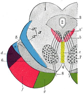
The midbrain or mesencephalon is the uppermost portion of the brainstem connecting the diencephalon and cerebrum with the pons. It consists of the cerebral peduncles, tegmentum, and tectum.

The raphe nuclei are a moderate-size cluster of nuclei found in the brain stem. They have 5-HT1 receptors which are coupled with Gi/Go-protein-inhibiting adenyl cyclase. They function as autoreceptors in the brain and decrease the release of serotonin. The anxiolytic drug Buspirone acts as partial agonist against these receptors. Selective serotonin reuptake inhibitor (SSRI) antidepressants are believed to act in these nuclei, as well as at their targets.

The solitary nucleus(SN) (nucleus of the solitary tract, nucleus solitarius, or nucleus tractus solitarii) is a series of neurons whose cell bodies form a roughly vertical column of grey matter in the medulla oblongata of the brainstem. Their axons form the bulk of the enclosed solitary tract. The solitary nucleus can be divided into different parts including dorsomedial, dorsolateral, and ventrolateral subnuclei.

The medial longitudinal fasciculus (MLF) is a prominent bundle of nerve fibres which pass within the ventral/anterior portion of periaqueductal gray of the mesencephalon (midbrain). It contains the interstitial nucleus of Cajal, responsible for oculomotor control, head posture, and vertical eye movement.

The reticular formation is a set of interconnected nuclei in the brainstem that spans from the lower end of the medulla oblongata to the upper end of the midbrain. The neurons of the reticular formation make up a complex set of neural networks in the core of the brainstem. The reticular formation is made up of a diffuse net-like formation of reticular nuclei which is not well-defined. It may be seen as being made up of all the interspersed cells in the brainstem between the more compact and named structures.

The pontine nuclei are all the neurons of the ventral pons. Corticopontine fibres project from the primary motor cortex to the ipsilateral pontine nucleus; pontocerebellar fibers then relay the information to the contralateral cerebellum via the middle cerebellar peduncle.

The fastigial nucleus is located in each cerebellar hemisphere. It is one of the four paired deep cerebellar nuclei of the cerebellum.

The flocculus is a small lobe of the cerebellum at the posterior border of the middle cerebellar peduncle anterior to the biventer lobule. Like other parts of the cerebellum, the flocculus is involved in motor control. It is an essential part of the vestibulo-ocular reflex, and aids in the learning of basic motor skills in the brain.
The dorsal longitudinal fasciculus (DLF) is a distinctive nerve tract in the midbrain. It extends from the hypothalamus rostrally to the spinal cord caudally, and contains both descending and ascending fibers.

The paramedian pontine reticular formation (PPRF) is a subset of neurons of the oral and caudal pontine reticular nuclei. With the abducens nucleus it makes up the horizontal gaze centre. It is situated in the pons adjacent to the abducens nucleus. It projects to the ipsilateral abducens nucleus, and contralateral oculomotor nucleus to mediate conjugate horizontal gaze and saccades.
The parvocellular reticular nucleus is part of the brain located dorsolateral to the caudal pontine reticular nucleus.
The caudal pontine reticular nucleus or nucleus reticularis pontis caudalis is a portion of the reticular formation, composed of gigantocellular neurons.
The gigantocellular reticular nucleus is the (efferent/motor) medial zone of the reticular formation of the caudal pons and rostral medulla oblongata. It consists of a substantial number of giant neurons, but also contains small and medium sized neurons.
The rostral interstitial nucleus of medial longitudinal fasciculus (riMLF) is a collection of neurons in the medial longitudinal fasciculus in the midbrain. It is responsible for mediating vertical conjugate eye movements and vertical saccades. It mostly projects efferents to the ipsilateral oculomotor and trochlear nuclei.
The nucleus prepositus or nucleus prepositus hypoglossi is one of the largest of the three perihypoglossal nuclei. It is situated in the caudal pons and rostral medulla oblongata. It contributes to several aspects of gaze control including the horizontal gaze holding system.
Serotonergic cell groups refer to collections of neurons in the central nervous system that have been demonstrated by histochemical fluorescence to contain the neurotransmitter serotonin (5-hydroxytryptamine). Since they are for the most part localized to classical brainstem nuclei, particularly the raphe nuclei, they are more often referred to by the names of those nuclei than by the B1-9 nomenclature. These cells appear to be common across most mammals and have two main regions in which they develop; one forms in the mesencephlon and the rostral pons and the other in the medulla oblongata and the caudal pons.
Perihypoglossal nuclei are three prominent groups of neurons in the caudal medulla oblongata near the hypoglossal nucleus: the nucleus prepositus hypoglossi, intercalated nucleus, and sublingual nucleus. They are involved in controlling eye movements: they send their principal projections to the three cranial nerve nuclei controlling extrinsic eye muscles via the medial longitudinal fasciculus.
In neuroanatomy, corticomesencephalic tract is a descending nerve tract that originates in the frontal eye field and terminates in the midbrain. Its fibers mediate conjugate eye movement.
The interstitial nucleus of Cajal is a collection of neurons in the mesencephalon (midbrain) which are involved in integrating eye position-velocity information in order to coordinate head-eye movements - especially those related to vertical and torsional conjugate eye movements (gaze). It also mediates vertical gaze holding.
References
- 1 2 Patestas, Maria A.; Gartner, Leslie P. (2016). A Textbook of Neuroanatomy (2nd ed.). Hoboken, New Jersey: Wiley-Blackwell. pp. 307–308, 310. ISBN 978-1-118-67746-9.
- ↑ Dergacheva OIu et al. Impulse activity of neurons in the nucleus pontis oralis in cats during sleep--wakefulness cycle. Ross Fiziol Zh Im I M Sechenova. 2002 Dec;88(12):1530-7.
| | This neuroanatomy article is a stub. You can help Wikipedia by expanding it. |