Median raphe nucleus

Last updated
This image shows the location of the raphe nucleus (seen in the bottom of the image near the brain stem) while also providing information about how it is used in the serotonin pathway which has various cognitive functions. The Median Raphe Nucleus is a part of the Raphe Nucleus. Pubmed equitativa hormonal.png
This image shows the location of the raphe nucleus (seen in the bottom of the image near the brain stem) while also providing information about how it is used in the serotonin pathway which has various cognitive functions. The Median Raphe Nucleus is a part of the Raphe Nucleus.
Median raphe nucleus
Details
Identifiers
Latin nucleus raphes medianus, nucleus centralis superior
NeuroNames 562
NeuroLex ID birnlex_889
TA98 A14.1.05.603
TA2 5956
FMA 72465
Anatomical terms of neuroanatomy

The median raphe nucleus (MRN or MnR), also known as the nucleus raphes medianus (NRM) [1] or superior central nucleus, is a brain region composed of polygonal, fusiform, and piriform neurons, which exists rostral to the nucleus raphes pontis. The MRN is located between the posterior end of the superior cerebellar peduncles and the V. Afferents of the motor nucleus. [2] It is one of two nuclei, the other being the dorsal raphe nucleus (DnR), in the midbrain-pons. [3]

Contents

The MRN projects extensively to the hippocampus, which is known to be essential for the formation of long-term memory. One recent study found that this raphe–hippocampus pathway plays a critical role in regulation of hippocampal activity and likely associated memory consolidation processes. It has also been found to play a role in anxiety and depression, as one of the few parts of the brain that creates tryptophan hydroxylase.

Description

The median raphe nucleus contains 5-hydroxytryptamine (serotonin, 5-HT) cell bodies that give rise to the majority of the ascending 5-HT projections to the forebrain limbic areas that control emotional behavior. [3] Because a dense population of neurons in the median raphe nucleus primarily contain serotonin, a prominent neurotransmitter in the median raphe nucleus is serotonin (5-HT). [4] Projections from the MRN extend to forebrain structures. [4] Distinct projection areas of the MnR innervates the medial septum, cingulate and dorsal hippocampus. [3] According to a study by McKenna and Vertes, around 8–12% of cells of the MnR were retrogradely double-labeled after paired injections in the Medial septum CA1 region, Medial Septum CA3 region, Medial Septum Dentate Gyrus of the dorsal hippocampus, the lateral Medial Septum Dentate Gyrus, and the Medial Septum ventral hippocampus. [5] These cells of the MnR that send collateral projections to the Medial Septum and hippocampus may serve a unique role in modulation of desynchronization of hippocampus EEG. [5] Also, the MnR has significantly more single- and double-labeled cells after paired injections to the various Medial Septum and hippocampus regions than in DnR which demonstrate that MnR has more stronger projections to the Medial Septum and hippocampus than the DnR. [5] MnR fibers are course and large with spherical varicosities. [3] Neurotoxic 5-HT-releasing agents selectively destroy DnR projection fibers without affecting the dense coarse fibers from the MnR. [6] Most of the fibers that distribute to the medial septum terminate selectively within the medial septum-vertical limb of the diagonal band nucleus (MS/DBv) and lateral aspects of lateral septum. [7] Most of the pronounced projections to hippocampal formation (HF) distribute to the stratum lacunosum-molecular of Ammon’s horn and granule cell layer and adjacent inner molecular layer of the dentate gyrus (DG). [7]

Projections stemming from the MRN modulate dopaminergic activity within the forebrain. [8] Additionally MnR projections are part of a behavioral disinhibition/inhibition system that produces phenotypes resembling behavioral variations manifested during manic and depressive phases of bipolar disorder. [8]

Inhibition of the MRN in cats by lysergic acid diethylamide (LSD) and psilocin, two serotonin agonist hallucinogens, leads to dose dependent behavioral changes, indicating the MRN may be an important site of action for humans hallucinations. [9]

The MRN projects extensively to the hippocampus, which is known to be essential for the formation of long-term memory. One recent study found that this raphe–hippocampus pathway plays a critical role in regulation of hippocampal activity and likely associated memory consolidation processes. [10] It has been shown that the MRN is a contributor of serotonergic agents, especially 5-HT to the hippocampus. These findings, together with the demonstration that serotonergic agents block Long Term Potentiation (LTP) and 5-HT antagonists enhance LTP and/or memory makes it clear that the MRN plays a part in formation of long term memory in the hippocampus. [5]

The MRN was found to play a vital role in hippocampal desynchronization; it exerts an inhibitory effect on the mechanism for hippocampal theta wave generation. [11] Also, median raphe nucleus suppresses theta bursts of the medial septal area neurons. [5] Numerous studies reveal that lesions in the MRN continuously caused ongoing theta activity, and when the MRN was injected with pharmacological agents, the neurons displayed inhibited activity or reduced excitatory to drive them to produce theta at short latencies and for long durations. [5] Therefore, MRN is a functional antagonist of the reticular formation which plays a critical role in hippocampal theta generation. [5]

Function

The MRN plays a role in the serotonin pathway. According to the study by Van De Kar and Lorens, it is the main source of 5-hydroxytryptamine (5-HT) to other parts of the brain [12] . 5-HT is another name for serotonin which is a neurotransmitter that is affected by many physical and emotional processes, including depression, mood, social functioning, exercise, and diet [13] . The MRN, when stimulated, significantly increases the amount of 5-HT present in the brain. This aided in forming the conclusion that the neurons in the MRN is the main contributor of 5-HT to the dorsal hippocampus as well as anterior and posterior cortical areas. [14] Furthermore, the MRN was found to an area in the brain that relates to inhibitory control by GABA of serotonin (5-HT). [15] The gamma-Aminobutyric acid (GABA) acts an inhibitory transmitter–when GABA antagonist were injected in the median raphe nucleus of rat, it was found that there was increase in serotonin turnover. [15] Such relationship is also seen in when the MRN is electrically stimulated and as a result behavioral inhibition is induced in rats. [16] These behaviors that are typically seen in rats during stressful situations involved crouching, teeth chattering, piloerection, and micturition. [16] When the MRN is electrically stimulated, the behavioral response was not only suppressed but there was a counteraction with para-chlorophenylalanine (PCPA), a serotonin synthesis inhibitor. [16] Such results demonstrate that the MRN is involved in behavioral inhibition as well.

Another function of the MRN is that it plays a role in depression. It has been discovered that the MRN is one of the few parts of the brain that creates tryptophan hydroxylase. Tryptophan hydroxylase is a rate-limiting enzyme that works with serotonin. When the levels of tryptophan hydroxylase 2 mRNA are elevated then more tryptophan hydroxylase is created. These elevated levels are associated with individuals who present as depressed suicides when compared to the nonpsychiatric controls. [17]  

Besides depression, MRN has been examined for its potential role in anxiety regulation. In examining the MRN, various animal models have shown that an inactivation of neurons containing the serotonin transmitter within the median raphe nucleus led to anxiolysis. [4] Anxiolysis refers to medications or drugs, for example, that results in a calm and relaxed state; it can be used to relieve anxiety or as a sedative. [18] Such relationship suggest that the MRN plays a regulatory function in anxiety. [4]

Research

The use of microdialysis and voltammetry in studies has indicated that neurotransmitter-mediated responses may be different, and chronic treatment with agonists may differentially regulate the MnR and DnR. [19] Results from these studies have demonstrated  the selective vulnerability of MnR or DnR. [19]

See also

Related Research Articles

<span class="mw-page-title-main">Neurotransmitter</span> Chemical substance that enables neurotransmission

A neurotransmitter is a signaling molecule secreted by a neuron to affect another cell across a synapse. The cell receiving the signal, or target cell, may be another neuron, but could also be a gland or muscle cell.

<span class="mw-page-title-main">Serotonin</span> Monoamine neurotransmitter

Serotonin or 5-hydroxytryptamine (5-HT) is a monoamine neurotransmitter. Its biological function is complex and multifaceted, modulating mood, cognition, reward, learning, memory, and numerous physiological processes such as vomiting and vasoconstriction. Approximately 90% of the serotonin the human body produces is in the gastrointestinal tract's enterochromaffin cells, where it regulates intestinal movements.

<span class="mw-page-title-main">Raphe nuclei</span>

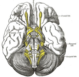
The raphe nuclei are a moderate-size cluster of nuclei found in the brain stem. They have 5-HT1 receptors which are coupled with Gi/Go-protein-inhibiting adenyl cyclase. They function as autoreceptors in the brain and decrease the release of serotonin. The anxiolytic drug Buspirone acts as partial agonist against these receptors. Selective serotonin reuptake inhibitor (SSRI) antidepressants are believed to act in these nuclei, as well as at their targets.

<span class="mw-page-title-main">Ventral tegmental area</span> Group of neurons on the floor of the midbrain

The ventral tegmental area (VTA), also known as the ventral tegmental area of Tsai, or simply ventral tegmentum, is a group of neurons located close to the midline on the floor of the midbrain. The VTA is the origin of the dopaminergic cell bodies of the mesocorticolimbic dopamine system and other dopamine pathways; it is widely implicated in the drug and natural reward circuitry of the brain. The VTA plays an important role in a number of processes, including reward cognition and orgasm, among others, as well as several psychiatric disorders. Neurons in the VTA project to numerous areas of the brain, ranging from the prefrontal cortex to the caudal brainstem and several regions in between.

In neuroanatomy, the pretectal area, or pretectum, is a midbrain structure composed of seven nuclei and comprises part of the subcortical visual system. Through reciprocal bilateral projections from the retina, it is involved primarily in mediating behavioral responses to acute changes in ambient light such as the pupillary light reflex, the optokinetic reflex, and temporary changes to the circadian rhythm. In addition to the pretectum's role in the visual system, the anterior pretectal nucleus has been found to mediate somatosensory and nociceptive information.

<span class="mw-page-title-main">Reticular formation</span> Spinal trigeminal nucleus

The reticular formation is a set of interconnected nuclei that are located throughout the brainstem. It is not anatomically well defined, because it includes neurons located in different parts of the brain. The neurons of the reticular formation make up a complex set of networks in the core of the brainstem that extend from the upper part of the midbrain to the lower part of the medulla oblongata. The reticular formation includes ascending pathways to the cortex in the ascending reticular activating system (ARAS) and descending pathways to the spinal cord via the reticulospinal tracts.

<span class="mw-page-title-main">Habenula</span>

In neuroanatomy, habenula originally denoted the stalk of the pineal gland, but gradually came to refer to a neighboring group of nerve cells with which the pineal gland was believed to be associated, the habenular nucleus. The habenular nucleus is a set of well-conserved structures in all vertebrate animals.

Theta waves generate the theta rhythm, a neural oscillation in the brain that underlies various aspects of cognition and behavior, including learning, memory, and spatial navigation in many animals. It can be recorded using various electrophysiological methods, such as electroencephalogram (EEG), recorded either from inside the brain or from electrodes attached to the scalp.

<span class="mw-page-title-main">Septal area</span> Area in the lower, posterior part of the medial surface of the frontal lobe

The septal area, consisting of the lateral septum and medial septum, is an area in the lower, posterior part of the medial surface of the frontal lobe, and refers to the nearby septum pellucidum.

<span class="mw-page-title-main">Dorsal raphe nucleus</span>

The dorsal raphe nucleus is located on the midline of the brainstem and is one of the raphe nuclei. It has rostral and caudal subdivisions.

<span class="mw-page-title-main">Nucleus raphe obscurus</span>

The nucleus raphe obscurus, despite the implications of its name, has some very specific functions and connections of afferent and efferent nature. The nucleus raphes obscurus projects to the cerebellar lobes VI and VII and to crus II along with the nucleus raphe pontis.

<span class="mw-page-title-main">Diagonal band of Broca</span>

The diagonal band of Broca is one of the basal forebrain structures that are derived from the ventral telencephalon during development. This structure forms the medial margin of the anterior perforated substance. This brain region was described by the French neuroanatomist Paul Broca.

<span class="mw-page-title-main">TPH2</span> Protein-coding gene in the species Homo sapiens

Tryptophan hydroxylase 2 (TPH2) is an isozyme of tryptophan hydroxylase found in vertebrates. In humans, TPH2 is primarily expressed in the serotonergic neurons of the brain, with the highest expression in the raphe nucleus of the midbrain. Until the discovery of TPH2 in 2003, serotonin levels in the central nervous system were believed to be regulated by serotonin synthesis in peripheral tissues, in which tryptophan hydroxylase is the dominant form.

<span class="mw-page-title-main">Hippocampus anatomy</span>

Hippocampus anatomy describes the physical aspects and properties of the hippocampus, a neural structure in the medial temporal lobe of the brain. It has a distinctive, curved shape that has been likened to the sea-horse monster of Greek mythology and the ram's horns of Amun in Egyptian mythology. This general layout holds across the full range of mammalian species, from hedgehog to human, although the details vary. For example, in the rat, the two hippocampi look similar to a pair of bananas, joined at the stems. In primate brains, including humans, the portion of the hippocampus near the base of the temporal lobe is much broader than the part at the top. Due to the three-dimensional curvature of this structure, two-dimensional sections such as shown are commonly seen. Neuroimaging pictures can show a number of different shapes, depending on the angle and location of the cut.

<span class="mw-page-title-main">Pallium (neuroanatomy)</span> Layers of grey and white matter that cover the upper surface of the cerebrum in vertebrates

In neuroanatomy, pallium refers to the layers of grey and white matter that cover the upper surface of the cerebrum in vertebrates. The non-pallial part of the telencephalon builds the subpallium. In basal vertebrates the pallium is a relatively simple three-layered structure, encompassing 3–4 histogenetically distinct domains, plus the olfactory bulb.

<span class="mw-page-title-main">Medial septal nucleus</span>

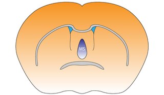
The medial septal nucleus (MS) is one of the septal nuclei. Neurons in this nucleus give rise to the bulk of efferents from the septal nuclei. A major projection from the medial septal nucleus terminates in the hippocampal formation.

Serotonergic cell groups refer to collections of neurons in the central nervous system that have been demonstrated by histochemical fluorescence to contain the neurotransmitter serotonin (5-hydroxytryptamine). Since they are for the most part localized to classical brainstem nuclei, particularly the raphe nuclei, they are more often referred to by the names of those nuclei than by the B1-9 nomenclature. These cells appear to be common across most mammals and have two main regions in which they develop; one forms in the mesencephlon and the rostral pons and the other in the medulla oblongata and the caudal pons.

<span class="mw-page-title-main">LP-44</span> Chemical compound

LP-44 is a drug which acts as a potent and selective agonist at the 5HT7 serotonin receptor. While LP-44 is less selective than the related compound LP-12, it has been more widely used in research and has been used to show the complex role of 5-HT7 receptors in several aspects of brain function, including regulation of the sleep-wake cycle and roles in stress, learning and memory.

The parabrachial nuclei, also known as the parabrachial complex, are a group of nuclei in the dorsolateral pons that surrounds the superior cerebellar peduncle as it enters the brainstem from the cerebellum. They are named from the Latin term for the superior cerebellar peduncle, the brachium conjunctivum. In the human brain, the expansion of the superior cerebellar peduncle expands the parabrachial nuclei, which form a thin strip of grey matter over most of the peduncle. The parabrachial nuclei are typically divided along the lines suggested by Baxter and Olszewski in humans, into a medial parabrachial nucleus and lateral parabrachial nucleus. These have in turn been subdivided into a dozen subnuclei: the superior, dorsal, ventral, internal, external and extreme lateral subnuclei; the lateral crescent and subparabrachial nucleus along the ventrolateral margin of the lateral parabrachial complex; and the medial and external medial subnuclei

References

  1. Federative Committee on Anatomical Terminology (FCAT) (1998). Terminologia Anatomica. Stuttgart: Thieme
  2. Walker, Emily P.; Tadi, Prasanna (2019), "Neuroanatomy, Nucleus Raphe", StatPearls, StatPearls Publishing, PMID   31335079 , retrieved 2019-09-24
  3. 1 2 3 4 Beck, Sheryl G.; Pan, Yu-Zhen; Akanwa, Adaure C.; Kirby, Lynn G. (February 2004). "Median and dorsal raphe neurons are not electrophysiologically identical". Journal of Neurophysiology. 91 (2): 994–1005. doi:10.1152/jn.00744.2003. ISSN   0022-3077. PMC   2830647 . PMID   14573555.
  4. 1 2 3 4 Andrade, Telma GCS; Zangrossi, Hélio; Graeff, Frederico G (2013-12-01). "The median raphe nucleus in anxiety revisited". Journal of Psychopharmacology. 27 (12): 1107–1115. doi:10.1177/0269881113499208. ISSN   0269-8811. PMID   23999409.
  5. 1 2 3 4 5 6 7 McKenna, James Timothy; Vertes, Robert P (April 2001). "Collateral projections from the median raphe nucleus to the medial septum and hippocampus". Brain Research Bulletin. 54 (6): 619–630. doi:10.1016/s0361-9230(01)00465-8. ISSN   0361-9230. PMID   11403988.
  6. Mamounas, L. A.; Mullen, C. A.; O'Hearn, E.; Molliver, M. E. (1991-12-15). "Dual serotoninergic projections to forebrain in the rat: morphologically distinct 5-HT axon terminals exhibit differential vulnerability to neurotoxic amphetamine derivatives". The Journal of Comparative Neurology. 314 (3): 558–586. doi:10.1002/cne.903140312. ISSN   0021-9967. PMID   1814975.
  7. 1 2 Vertes, Robert P.; Fortin, William J.; Crane, Alison M. (1999). "Projections of the median raphe nucleus in the rat". Journal of Comparative Neurology. 407 (4): 555–582. doi:10.1002/(sici)1096-9861(19990517)407:4<555::aid-cne7>3.0.co;2-e. ISSN   1096-9861. PMID   10235645.
  8. 1 2 Pezzato, Fernanda A.; Can, Adem; Hoshino, Katsumasa; Horta, José de Anchieta C.; Mijares, Miriam G.; Gould, Todd D. (2015-04-01). "Effect of lithium on behavioral disinhibition induced by electrolytic lesion of the median raphe nucleus". Psychopharmacology. 232 (8): 1441–1450. doi:10.1007/s00213-014-3775-z. ISSN   1432-2072. PMC   4388762 . PMID   25345734.
  9. Trulson, M.E., Preussler DW and Trulson V.M. Differential effects of hallucinogenic drugs on the activity of serotonin-containing neurons in the nucleus centralis superior and nucleus raphe pallidus in free-moving cats. American Society for Pharmacology and Experimental Therapeutics Volume 228, Issue 1, pp. 94-102, 1 January 1984
  10. 4. Wang, D.V., Yau, H., Broker, C.J., Tsou, J., Bonci, A. & Ikemoto, S. Mesopontine median raphe regulates hippocampal ripple oscillation and memory consolidation. Nature Neuroscience 18, 728-735, 2015
  11. Maru, Eiichi; Takahashi, Lorey K.; Iwahara, Shinkuro (1979-03-16). "Effects of median raphe nucleus lesions on hippocampal EEG in the freely moving rat". Brain Research. 163 (2): 223–234. doi:10.1016/0006-8993(79)90351-2. ISSN   0006-8993. PMID   218681.
  12. Van De Kar, L. D.; Lorens, S. A. (1979-02-16). "Differential serotonergic innervation of individual hypothalamic nuclei and other forebrain regions by the dorsal and median midbrain raphe nuclei". Brain Research. 162 (1): 45–54. doi:10.1016/0006-8993(79)90754-6. ISSN   0006-8993. PMID   761086.
  13. Young, Simon N. (November 2007). "How to increase serotonin in the human brain without drugs". Journal of Psychiatry & Neuroscience. 32 (6): 394–399. ISSN   1180-4882. PMC   2077351 . PMID   18043762.
  14. McQuade, R.; Sharp, T. (1997). "Functional Mapping of Dorsal and Median Raphe 5-Hydroxytryptamine Pathways in Forebrain of the Rat Using Microdialysis". Journal of Neurochemistry. 69 (2): 791–796. doi: 10.1046/j.1471-4159.1997.69020791.x . ISSN   1471-4159. PMID   9231740.
  15. 1 2 Forchetti, Concetta M.; Meek, James L. (1981-02-09). "Evidence for a tonic GABAergic control of serotonin neurons in the median raphe nucleus". Brain Research. 206 (1): 208–212. doi:10.1016/0006-8993(81)90118-9. ISSN   0006-8993. PMID   7470888.
  16. 1 2 3 Graeff, F. G.; Silveira Filho, N. G. (1978-10-01). "Behavioral inhibition induced by electrical stimulation of the median raphe nucleus of the rat". Physiology & Behavior. 21 (4): 477–484. doi:10.1016/0031-9384(78)90116-6. ISSN   0031-9384. PMID   154108.
  17. Bach-Mizrachi, Helene; Underwood, Mark D.; Kassir, Suham A.; Bakalian, Mihran J.; Sibille, Etienne; Tamir, Hadassah; Mann, J. John; Arango, Victoria (April 2006). "Neuronal Tryptophan Hydroxylase mRNA Expression in the Human Dorsal and Median Raphe Nuclei: Major Depression and Suicide". Neuropsychopharmacology. 31 (4): 814–824. doi: 10.1038/sj.npp.1300897 . ISSN   1740-634X. PMID   16192985.
  18. "NCI Dictionary of Cancer Terms". National Cancer Institute. 2011-02-02. Retrieved 2019-11-02.
  19. 1 2 Kreiss, D. S.; Lucki, I. (February 1997). "Chronic administration of the 5-HT1A receptor agonist 8-OH-DPAT differentially desensitizes 5-HT1A autoreceptors of the dorsal and median raphe nuclei". Synapse (New York, N.Y.). 25 (2): 107–116. doi:10.1002/(SICI)1098-2396(199702)25:2<107::AID-SYN1>3.0.CO;2-G. ISSN   0887-4476. PMID   9021891.