Cochlear nucleus

Last updated
Cochlear nuclei
Gray691.png
Dissection of brainstem. Dorsal view. ("Cochlear nucleus" is labeled on left, fifth from the bottom.)
Gray760.png
Terminal nuclei of the cochlear nerve, with their upper connections. (Schematic.) The vestibular nerve with its terminal nuclei and their efferent fibers have been suppressed. On the other hand, in order not to obscure the trapezoid body, the efferent fibers of the terminal nuclei on the right side have been resected in a considerable portion of their extent. The trapezoid body, therefore, shows only one-half of its fibers, viz., those that come from the left.
  1. Vestibular nerve, divided at its entrance into the medulla oblongata
  2. Cochlear nerve
  3. Accessory nucleus of acoustic nerve
  4. Tuberculum acusticum
  5. Efferent fibers of accessory nucleus
  6. Efferent fibers of tuberculum acusticum, forming the striae medullares, with 6’, their direct bundle going to the superior olivary nucleus of the same side; 6’’, their decussating bundles going to the superior olivary nucleus of the opposite side
  7. Superior olivary nucleus
  8. Trapezoid body
  9. Trapezoid nucleus
  10. Central acoustic tract (lateral lemniscus)
  11. Raphé
  12. Pyramidal tracts
  13. Fourth ventricle
  14. Inferior peduncle
Details
Part of brainstem
System Auditory system
Artery AICA
Identifiers
Latin nuclei cochleares
MeSH D017626
NeuroNames 720
NeuroLex ID birnlex_1151
TA98 A14.1.04.247
A14.1.05.430
TA2 6006, 6007
FMA 72240
Anatomical terms of neuroanatomy

The cochlear nuclear (CN) complex comprises two cranial nerve nuclei in the human brainstem, the ventral cochlear nucleus (VCN) and the dorsal cochlear nucleus (DCN). The ventral cochlear nucleus is unlayered whereas the dorsal cochlear nucleus is layered. Auditory nerve fibers, fibers that travel through the auditory nerve (also known as the cochlear nerve or eighth cranial nerve) carry information from the inner ear, the cochlea, on the same side of the head, to the nerve root in the ventral cochlear nucleus. At the nerve root the fibers branch to innervate the ventral cochlear nucleus and the deep layer of the dorsal cochlear nucleus. All acoustic information thus enters the brain through the cochlear nuclei, where the processing of acoustic information begins. The outputs from the cochlear nuclei are received in higher regions of the auditory brainstem.

Contents

Structure

The cochlear nuclei (CN) are located at the dorso-lateral side of the brainstem, spanning the junction of the pons and medulla.

Projections to the cochlear nuclei

The major input to the cochlear nucleus is from the auditory nerve, a part of cranial nerve VIII (the vestibulocochlear nerve). The auditory nerve fibers form a highly organized system of connections according to their peripheral innervation of the cochlea. Axons from the spiral ganglion cells of the lower frequencies innervate the ventrolateral portions of the ventral cochlear nucleus and lateral-ventral portions of the dorsal cochlear nucleus. The axons from the higher frequency organ of corti hair cells project to the dorsal portion of the ventral cochlear nucleus and the dorsal-medial portions of the dorsal cochlear nucleus. The mid frequency projections end up in between the two extremes; in this way the tonotopic organization that is established in the cochlea is preserved in the cochlear nuclei. This tonotopic organization is preserved because only a few inner hair cells synapse on the dendrites of a nerve cell in the spiral ganglion, and the axon from that nerve cell synapses on only a very few dendrites in the cochlear nucleus. In contrast with the VCN that receives all acoustic input from the auditory nerve, the DCN receives input not only from the auditory nerve but it also receives acoustic input from neurons in the VCN (T stellate cells). The DCN is therefore in a sense a second order sensory nucleus.

The cochlear nuclei have long been thought to receive input only from the ipsilateral ear. There is evidence, however, for stimulation from the contralateral ear via the contralateral CN, [2] and also the somatosensory parts of the brain. [3]

Projections from the cochlear nuclei

There are three major fiber bundles, axons of cochlear nuclear neurons, that carry information from the cochlear nuclei to targets that are mainly on the opposite side of the brain. Through the medulla, one projection goes to the contralateral superior olivary complex (SOC) via the trapezoid body, whilst the other half shoots to the ipsilateral SOC. This pathway is called the ventral acoustic stria (VAS or, more commonly, the trapezoid body). Another pathway, called the dorsal acoustic stria (DAS, also known as the stria of von Monakow), rises above the medulla into the pons where it hits the nuclei of the lateral lemniscus along with its kin, the intermediate acoustic stria (IAS, also known as the stria of Held). The IAS decussates across the medulla, before joining the ascending fibers in the contralateral lateral lemniscus. The lateral lemniscus contains cells of the nuclei of the lateral lemniscus, and in turn projects to the inferior colliculus. The inferior colliculus receives direct, monosynaptic projections from the superior olivary complex, the contralateral dorsal acoustic stria, some classes of stellate neurons of the VCN, as well as from the different nuclei of the lateral lemniscus.

Most of these inputs terminate in the inferior colliculus, although there are a few small projections that bypass the inferior colliculus and project to the medial geniculate, or other forebrain structures.

Histology

Three types of principal cells convey information out of the ventral cochlear nucleus: Bushy cells, stellate cells, and octopus cells.

Two types of principal cells convey information out of the dorsal cochlear nucleus (DCN) to the contralateral inferior colliculus. The principal cells receive two systems of inputs. Acoustic input comes to the deep layer through several paths. Excitatory acoustic input comes from auditory nerve fibers and also from stellate cells of the VCN. Acoustic input is also conveyed through inhibitory interneurons (tuberculoventral cells of the DCN and "wide band inhibitors" in the VCN). Through the outermost molecular layer, the DCN receives other types of sensory information, most importantly information about the location of the head and ears, through parallel fibers. This information is distributed through a cerebellar like circuit that also includes inhibitory interneurons.

Function

The cochlear nuclear complex is the first integrative, or processing, stage in the auditory system. Information is brought to the nuclei from the ipsilateral cochlea via the cochlear nerve. Several tasks are performed in the cochlear nuclei. By distributing acoustic input to multiple types of principal cells, the auditory pathway is subdivided into parallel ascending pathways, which can simultaneously extract different types of information. The cells of the ventral cochlear nucleus extract information that is carried by the auditory nerve in the timing of firing and in the pattern of activation of the population of auditory nerve fibers. The cells of the dorsal cochlear nucleus perform a non-linear spectral analysis and place that spectral analysis into the context of the location of the head, ears and shoulders and that separate expected, self-generated spectral cues from more interesting, unexpected spectral cues using input from the auditory cortex, pontine nuclei, trigeminal ganglion and nucleus, dorsal column nuclei and the second dorsal root ganglion. It is likely that these neurons help mammals to use spectral cues for orienting toward those sounds. The information is used by higher brainstem regions to achieve further computational objectives (such as sound source location or improvement in signal-to-noise ratio). The inputs from these other areas of the brain probably play a role in sound localization.

In order to understand in more detail the specific functions of the cochlear nuclei it is first necessary to understand the way sound information is represented by the fibers of the auditory nerve. Briefly, there are around 30,000 auditory nerve fibres in each of the two auditory nerves. Each fiber is an axon of a spiral ganglion cell that represents a particular frequency of sound, and a particular range of loudness. Information in each nerve fibre is represented by the rate of action potentials as well as the particular timing of individual action potentials. The particular physiology and morphology of each cochlear nucleus cell type enhances different aspects of sound information.

See also

Additional images

Related Research Articles

Articles related to anatomy include:

<span class="mw-page-title-main">Brainstem</span> Posterior part of the brain, adjoining and structurally continuous

The brainstem is the stalk-like part of the brain that interconnects the cerebrum and diencephalon with the spinal cord. In the human brain, the brainstem is composed of the midbrain, the pons, and the medulla oblongata. The midbrain is continuous with the thalamus of the diencephalon through the tentorial notch.

<span class="mw-page-title-main">Midbrain</span> Forward-most portion of the brainstem

The midbrain or mesencephalon is the rostral-most portion of the brainstem connecting the diencephalon and cerebrum with the pons. It consists of the cerebral peduncles, tegmentum, and tectum.

<span class="mw-page-title-main">Spinothalamic tract</span> Sensory pathway from the skin to the thalamus

The spinothalamic tract is a part of the anterolateral system or the ventrolateral system, a sensory pathway to the thalamus. From the ventral posterolateral nucleus in the thalamus, sensory information is relayed upward to the somatosensory cortex of the postcentral gyrus.

<span class="mw-page-title-main">Dorsal column–medial lemniscus pathway</span> Sensory spinal pathway

The dorsal column–medial lemniscus pathway (DCML) is a sensory pathway of the central nervous system that conveys sensations of fine touch, vibration, two-point discrimination, and proprioception from the skin and joints. It transmits information from the body to the primary somatosensory cortex in the postcentral gyrus of the parietal lobe of the brain. The pathway receives information from sensory receptors throughout the body, and carries this in nerve tracts in the white matter of the dorsal column of the spinal cord to the medulla, where it is continued in the medial lemniscus, on to the thalamus and relayed from there through the internal capsule and transmitted to the somatosensory cortex. The name dorsal-column medial lemniscus comes from the two structures that carry the sensory information: the dorsal columns of the spinal cord, and the medial lemniscus in the brainstem.

<span class="mw-page-title-main">Auditory system</span> Sensory system used for hearing

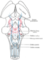
The auditory system is the sensory system for the sense of hearing. It includes both the sensory organs and the auditory parts of the sensory system.

<span class="mw-page-title-main">Lateral lemniscus</span>

The lateral lemniscus is a tract of axons in the brainstem that carries information about sound from the cochlear nucleus to various brainstem nuclei and ultimately the contralateral inferior colliculus of the midbrain. Three distinct, primarily inhibitory, cellular groups are located interspersed within these fibers, and are thus named the nuclei of the lateral lemniscus.

<span class="mw-page-title-main">Inferior colliculus</span> Midbrain structure involved in the auditory pathway

The inferior colliculus (IC) is the principal midbrain nucleus of the auditory pathway and receives input from several peripheral brainstem nuclei in the auditory pathway, as well as inputs from the auditory cortex. The inferior colliculus has three subdivisions: the central nucleus, a dorsal cortex by which it is surrounded, and an external cortex which is located laterally. Its bimodal neurons are implicated in auditory-somatosensory interaction, receiving projections from somatosensory nuclei. This multisensory integration may underlie a filtering of self-effected sounds from vocalization, chewing, or respiration activities.

<span class="mw-page-title-main">Trapezoid body</span> Part of the auditory pathway

The trapezoid body is part of the auditory pathway where some of the axons coming from the cochlear nucleus decussate to the other side before traveling on to the superior olivary nucleus. This is believed to help with localization of sound.

<span class="mw-page-title-main">Spinocerebellar tract</span> Nerve tract in humans

The spinocerebellar tract is a nerve tract originating in the spinal cord and terminating in the same side (ipsilateral) of the cerebellum.

<span class="mw-page-title-main">Medial geniculate nucleus</span>

The medial geniculate nucleus (MGN) or medial geniculate body (MGB) is part of the auditory thalamus and represents the thalamic relay between the inferior colliculus (IC) and the auditory cortex (AC). It is made up of a number of sub-nuclei that are distinguished by their neuronal morphology and density, by their afferent and efferent connections, and by the coding properties of their neurons. It is thought that the MGN influences the direction and maintenance of attention.

<span class="mw-page-title-main">Cochlear nerve</span> Nerve carrying auditory information from the inner ear to the brain

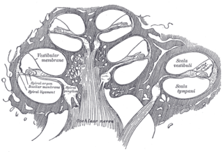
The cochlear nerve is one of two parts of the vestibulocochlear nerve, a cranial nerve present in amniotes, the other part being the vestibular nerve. The cochlear nerve carries auditory sensory information from the cochlea of the inner ear directly to the brain. The other portion of the vestibulocochlear nerve is the vestibular nerve, which carries spatial orientation information to the brain from the semicircular canals, also known as semicircular ducts.

<span class="mw-page-title-main">Facial motor nucleus</span>

The facial motor nucleus is a collection of neurons in the brainstem that belong to the facial nerve. These lower motor neurons innervate the muscles of facial expression and the stapedius.

<span class="mw-page-title-main">Dorsal cochlear nucleus</span>

The dorsal cochlear nucleus is a cortex-like structure on the dorso-lateral surface of the brainstem. Along with the ventral cochlear nucleus (VCN), it forms the cochlear nucleus (CN), where all auditory nerve fibers from the cochlea form their first synapses.

<span class="mw-page-title-main">Superior olivary complex</span> Collection of brainstem nuclei related to hearing

The superior olivary complex (SOC) or superior olive is a collection of brainstem nuclei that is located in pons, functions in multiple aspects of hearing and is an important component of the ascending and descending auditory pathways of the auditory system. The SOC is intimately related to the trapezoid body: most of the cell groups of the SOC are dorsal to this axon bundle while a number of cell groups are embedded in the trapezoid body. Overall, the SOC displays a significant interspecies variation, being largest in bats and rodents and smaller in primates.

<span class="mw-page-title-main">Interaural time difference</span> Difference in time that it takes a sound to travel between two ears

The interaural time difference when concerning humans or animals, is the difference in arrival time of a sound between two ears. It is important in the localization of sounds, as it provides a cue to the direction or angle of the sound source from the head. If a signal arrives at the head from one side, the signal has further to travel to reach the far ear than the near ear. This pathlength difference results in a time difference between the sound's arrivals at the ears, which is detected and aids the process of identifying the direction of sound source.

Binaural fusion or binaural integration is a cognitive process that involves the combination of different auditory information presented binaurally, or to each ear. In humans, this process is essential in understanding speech as one ear may pick up more information about the speech stimuli than the other.

<span class="mw-page-title-main">Ventral cochlear nucleus</span>

In the ventral cochlear nucleus (VCN), auditory nerve fibers enter the brain via the nerve root in the VCN. The ventral cochlear nucleus is divided into the anterior ventral (anteroventral) cochlear nucleus (AVCN) and the posterior ventral (posteroventral) cochlear nucleus (PVCN). In the VCN, auditory nerve fibers bifurcate, the ascending branch innervates the AVCN and the descending branch innervates the PVCN and then continue to the dorsal cochlear nucleus. The orderly innervation by auditory nerve fibers gives the AVCN a tonotopic organization along the dorsoventral axis. Fibers that carry information from the apex of the cochlea that are tuned to low frequencies contact neurons in the ventral part of the AVCN; those that carry information from the base of the cochlea that are tuned to high frequencies contact neurons in the dorsal part of the AVCN. Several populations of neurons populate the AVCN. Bushy cells receive input from auditory nerve fibers through particularly large endings called end bulbs of Held. They contact stellate cells through more conventional boutons.

<span class="mw-page-title-main">Calyx of Held</span>

The Calyx of Held is a particularly large synapse in the mammalian auditory central nervous system, so named after Hans Held who first described it in his 1893 article Die centrale Gehörleitung because of its resemblance to the calyx of a flower. Globular bushy cells in the anteroventral cochlear nucleus (AVCN) send axons to the contralateral medial nucleus of the trapezoid body (MNTB), where they synapse via these calyces on MNTB principal cells. These principal cells then project to the ipsilateral lateral superior olive (LSO), where they inhibit postsynaptic neurons and provide a basis for interaural level detection (ILD), required for high frequency sound localization. This synapse has been described as the largest in the brain.

References

PD-icon.svgThis article incorporates text in the public domain from page 788 of the 20th edition of Gray's Anatomy (1918)Young ED, Spirou GA, Rice JJ, Voigt HF (June 1992). "Neural organization and responses to complex stimuli in the dorsal cochlear nucleus". Philos. Trans. R. Soc. Lond. B Biol. Sci. 336 (1278): 407–13. doi:10.1098/rstb.1992.0076. PMID   1354382.

  1. Middlebrooks, J.C. (2009). "Auditory System: Central Pathways". In Squire (ed.). Encyclopedia of Neuroscience. Academic Press. pp. 745–752, here: p. 745 f.
  2. Davis KA (September 2005). "Contralateral effects and binaural interactions in dorsal cochlear nucleus". J. Assoc. Res. Otolaryngol. 6 (3): 280–96. doi:10.1007/s10162-005-0008-5. PMC   2504593 . PMID   16075189.
  3. Shore, S.E. (2009). "Auditory/Somatosensory Interactions". In Squire (ed.). Encyclopedia of Neuroscience. Academic Press. pp. 691–5.